 Back
BackTissue Level of Organization: Epithelial and Connective Tissues
Study Guide - Smart Notes
 Tailored notes based on your materials, expanded with key definitions, examples, and context.
Tissue Level of Organization
Introduction to Tissues
Tissues are collections of similar cells and cell products that perform specific functions in the body. The study of tissues is known as histology. Cells within tissues are structured differently, resulting in diverse functions. Tissues combine to form organs, which carry out complex physiological tasks.
Definition: Tissue is a group of similar cells working together to perform a specific function.
Histology: The microscopic study of tissue structure and function.
Organization: Cells & extracellular material form tissues, which then form organs.
Four Major Tissue Types
The human body contains four primary tissue types, each with distinct roles:
Epithelial Tissue: Covers exposed surfaces, lines internal passageways, and forms glands.
Connective Tissue: Fills internal spaces, supports other tissues, transports materials, and stores energy.
Muscle Tissue: Specialized for contraction, producing movement.
Neural Tissue: Conducts electrical impulses, carrying information throughout the body.
Epithelial Tissue
Components and Functions
Epithelial tissue consists of epithelia (layers of cells covering surfaces) and glands (structures producing secretions). Epithelia cover exposed surfaces and line internal cavities, often containing secretory cells. Glands are derived from epithelia and are classified as exocrine (secrete onto surfaces or into ducts) or endocrine (secrete hormones into interstitial fluid and blood).
Physical Protection: Guards against abrasion, dehydration, and chemical/biological agents.
Control Permeability: Regulates absorption and filtration of substances.
Provide Sensation: Contains sensory nerve endings.
Produce Secretions: Forms glands for secretion of substances.
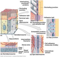
Intercellular Junctions
Epithelial cells are tightly bound by specialized junctions:
Occluding (Tight) Junctions: Prevent passage of water and solutes between cells.
Gap Junctions: Allow diffusion of ions and small molecules between cells.
Desmosomes: Provide resistance to stretching and twisting.
Basement Membrane
Epithelial tissue is avascular and anchored to a basement membrane, which attaches cells to underlying structures. The basement membrane consists of two layers:
Basal Lamina: Attaches to epithelium.
Reticular Lamina: Deeper layer, closer to the body.
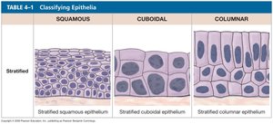
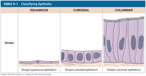
Classification of Epithelial Tissue
Epithelial tissues are classified by cell shape and number of layers:
Squamous: Flat, oval cells.
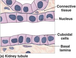
Cuboidal: Cube-shaped cells with centrally located nuclei.
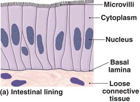
Columnar: Elongated, column-shaped cells with nuclei near the basal lamina.
Glandular: Specialized for secretion.
Organization:
Simple: One layer of cells, all touching the basal lamina.
Stratified: Multiple layers, not all touching the basal lamina.
Types of Epithelial Tissue
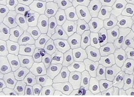
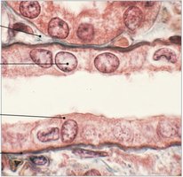
Simple Squamous Epithelium
Simple squamous consists of a single layer of flat cells, facilitating gas exchange and diffusion.
Locations: Lines blood and lymph vessels, alveoli of lungs, body cavities, portions of kidney tubules.
Special Names: Endothelium (lining heart and blood vessels), Mesothelium (lining body cavities).
Simple Cuboidal Epithelium
Composed of cube-shaped cells with centrally located nuclei, this tissue is specialized for secretion and absorption.
Locations: Kidneys, glands, ducts, thyroid gland.
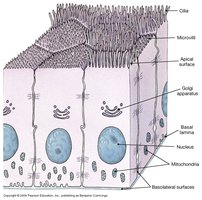
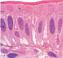
Simple Columnar Epithelium
Consists of elongated cells with nuclei near the basal lamina, often containing goblet cells for mucus secretion.
Functions: Secretion of digestive fluids, absorption of nutrients, protection.
Locations: Digestive system, uterine tubes, gallbladder.
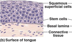
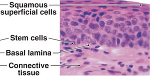
Stratified Squamous Epithelium
Multiple layers of flattened cells provide protection against abrasion.
Locations: Skin, mouth, throat, vagina, anal canal.
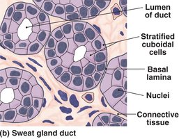
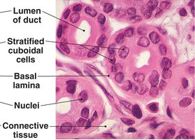
Stratified Cuboidal Epithelium
Composed of 2-3 layers of cuboid cells, this tissue provides protection.
Locations: Ducts of mammary glands, sweat glands.
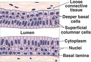
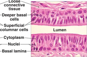
Stratified Columnar Epithelium
Several layers of cells, with elongated cells along the lumen, provide protection.
Locations: Urethra, anus, pharynx.
Transitional Epithelium
Several layers of cells with curved apical surfaces tolerate repeated cycles of stretching and recoiling.
Function: Prevents urine from moving backward.
Locations: Urinary bladder, ureters.
Pseudostratified Ciliated Columnar Epithelium
Appears layered, but all cells touch the basal lamina; contains cilia and goblet cells.
Functions: Moves mucus, provides protection, secretion.
Locations: Nasal cavity, trachea, bronchi.
Glandular Epithelium
Exocrine Glands: Secrete products into ducts (e.g., sweat, salivary, mammary glands).
Endocrine Glands: Secrete hormones into body fluids or blood (e.g., pancreas, adrenal glands).
Types of Secretion: Serous (watery), mucous (thicker), mixed (both).
Modes of Secretion: Merocrine (exocytosis), Apocrine (portion of cytoplasm released), Holocrine (cell dies during secretion).
Connective Tissue
Functions and Characteristics
Connective tissue binds, supports, protects, serves as frameworks, fills spaces, stores fat, produces blood cells, protects against infection, repairs tissue damage, and transports materials.
Extracellular Matrix: Composed of protein fibers and ground substance, determining tissue function.
Blood Supply: Generally good, except in cartilage.
Types of Connective Tissue
Loose Connective Tissue: Areolar, adipose, reticular.
Dense Connective Tissue: Regular, irregular, elastic.
Cartilage: Hyaline, elastic, fibrocartilage.
Other: Blood, lymph, bone.
Connective Tissue Fiber Types
Collagenous Fibers: Strong, flexible, most common.
Elastic Fibers: Stretch and recoil.
Reticular Fibers: Thin, form supportive networks.
Major Cell Types
Fibroblasts: Secrete fibers.
Fibrocytes: Maintain fibers.
Macrophages/Microphages: Defend against infection.
Adipocytes: Store fat.
Mesenchyme Cells: Stem cells for connective tissue.
Melanocytes: Produce melanin.
Mast Cells: Release heparin and histamine.
Lymphocytes: Immune defense.
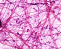
Areolar Connective Tissue
Extremely loose, open organization of cells, supports skin and binds body parts together.
Location: Under skin.
Function: Allows for independent movement, distorts and returns to original shape.
Adipose Tissue
Stores fat in vacuoles, cushions organs, and provides insulation.
Locations: Skin, buttocks, breasts, around eyes.
Reticular Tissue
Forms a complex, 3-D framework for organs such as liver, bone marrow, spleen, and lymph nodes.
Dense Regular Connective Tissue
Tightly packed fibers for attachment.
Locations: Tendons, ligaments, aponeuroses.
Elastic Tissue
Stabilizes, cushions, and permits expansion; dominated by elastic fibers.
Location: Vertebrae.
Dense Irregular Connective Tissue
Interwoven network provides strength and support.
Locations: Capsules, covering of bones.
Blood and Lymph
Blood: Transports substances, defends body, clots blood. Contains erythrocytes (gas transport), leukocytes (infection defense), and thrombocytes (clotting).
Lymph: Forms as extracellular fluid, purified by lymph nodes, returns to venous system, monitored by immune system.
Cartilage
Chondrocytes: Cartilage cells in lacunae.
Perichondrium: Covering surrounding cartilage.
Types: Hyaline (reduces friction), Elastic (supportive, bends easily), Fibrocartilage (limits movement, prevents bone contact).
Bone
Most rigid tissue, matrix hardened by calcium.
Osteocytes: Bone cells in lacunae.
Functions: Protection, blood cell production.
Muscle and Nervous Tissues
Muscle Tissue
Specialized for contraction, producing movement.
Skeletal Muscle: Voluntary, striated, multinucleated, attached to bones.
Cardiac Muscle: Involuntary, striated, branched, single nucleus, found in heart.
Smooth Muscle: Involuntary, non-striated, single nucleus, found in hollow organs.
Nervous Tissue
Conducts electrical impulses, rapidly senses environment, processes information, and controls responses.
Neuron: Transmits electrical signals.
Neuroglia: Supporting cells, repair and nourish neurons.
Membranes
Types of Membranes
Physical barriers that line or cover portions of the body, consisting of epithelium supported by connective tissue.
Mucous Membranes: Line passageways with external openings; moist surfaces facilitate absorption and excretion.
Serous Membranes: Line cavities not open to the outside; contain serous fluid to reduce friction.
Cutaneous Membrane: Skin; thick, waterproof, dry.
Synovial Membranes: Line joint cavities; produce synovial fluid for lubrication.
