 Back
BackTissue Level of Organization: Epithelial, Connective, Muscle, and Nervous Tissues
Study Guide - Smart Notes
 Tailored notes based on your materials, expanded with key definitions, examples, and context.
Tissue Level of Organization
Overview of Body Organization
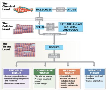
The human body is organized into hierarchical levels, starting from atoms and molecules, progressing to cells, and then to tissues. Tissues are groups of cells working together to perform specific functions. The study of tissues is known as histology. There are four basic types of tissues: epithelial, connective, muscle, and nervous.
Microscopy and Studying Tissues
Microscopy is essential for studying tissues at the cellular and subcellular levels. There are several types of microscopes:
Light Microscope (LM): Uses visible light, suitable for general cell structures and tissues. Magnification up to ~2000×.
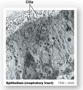
Transmission Electron Microscope (TEM): Uses electron beams through specimens, ideal for internal ultrastructure. Magnification up to ~1,000,000×.
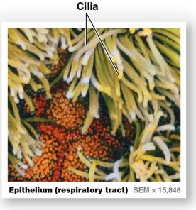
Scanning Electron Microscope (SEM): Uses electron beams for surface scanning, best for surface details and texture. Magnification up to ~100,000×.

Epithelial Tissue
Characteristics and Functions
Epithelial tissue covers exposed surfaces, lines internal passageways and chambers, and forms secretory glands. It is avascular, meaning it lacks blood vessels, and relies on diffusion for nutrient supply. Epithelial tissue functions include:
Physical protection: Protects surfaces from abrasion, dehydration, and destruction.
Control permeability: Selective absorption and secretion.
Provide sensation: Detects environmental changes.
Produce specialized secretions: Glandular cells produce sweat, oils, and other secretions.


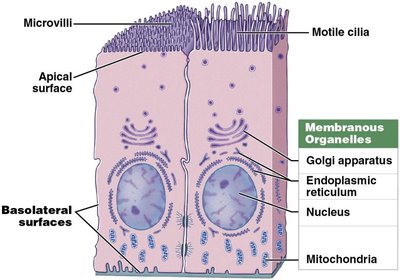
Structure of Epithelial Tissue
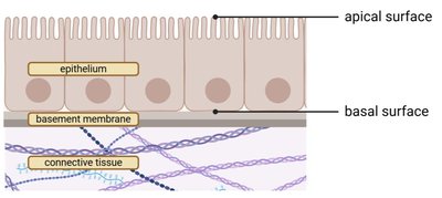
Epithelial cells have distinct surfaces:
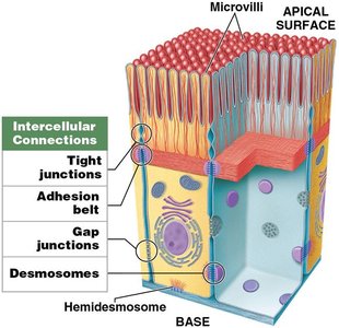
Apical surface: Faces the exterior or internal space; may have microvilli (for absorption) or cilia (for movement).
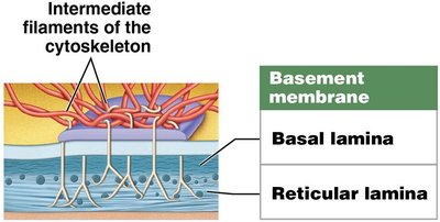
Basal surface: Attached to underlying connective tissue via the basement membrane.
Basolateral surface: Includes base and sides attached to neighboring cells.


Classification of Epithelial Tissue
Epithelial tissues are classified by cell layers and cell shape:
Simple epithelia: Single layer of cells; found in areas where absorption, diffusion, or filtration occurs.
Stratified epithelia: Multiple layers; found in areas needing protection from abrasion or chemical stress.

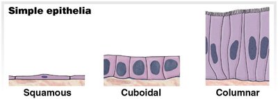


Types of Epithelial Cells
Squamous: Thin and flat; found in areas of absorption and diffusion.
Cuboidal: Cube-shaped; found in glands and ducts.
Columnar: Taller than wide; found in areas of absorption and secretion.
Glandular Epithelia
Glandular epithelium is specialized for secretion. Glands are classified as:
Endocrine: Secrete hormones directly into blood or tissue fluid; no ducts.
Exocrine: Secrete products onto surfaces or into ducts.
Methods of Secretion
Merocrine: Product released by exocytosis; cell remains intact.
Apocrine: Product released with part of cell cytoplasm; apical portion pinches off.
Holocrine: Entire cell bursts, releasing product and killing cell; replaced by stem cells.
Structural Classification
Simple: Single duct, does not divide.
Compound: Duct divides one or more times.
Tubular: Cells form tubes.
Alveolar/Acinar: Cells form sacs.
Tubuloalveolar: Cells form both tubes and sacs.
Connective Tissue
Characteristics and Functions
Connective tissue supports, binds, and transports materials throughout the body. It contains fewer cells but abundant extracellular matrix (ECM), which consists of fibers and ground substance. Functions include:
Structural framework
Transport of fluids and dissolved materials
Protection of organs
Support and interconnection of tissues
Energy storage (triglycerides)
Defense against microorganisms
Components of Connective Tissue
Specialized cells
Extracellular protein fibers
Ground substance (fluid)
Types of Connective Tissue
Connective tissue proper: Loose (areolar, adipose, reticular) and dense (regular, irregular, elastic).
Fluid connective tissue: Blood and lymph.
Supporting connective tissue: Cartilage and bone.
Loose Connective Tissue Proper
Areolar: Packing material; under skin, around vessels and organs.
Adipose: Fat storage, insulation, cushioning; under skin, around eyes and kidneys.
Reticular: Support for soft organs; lymph nodes, spleen, bone marrow, liver.
Dense Connective Tissue Proper
Dense regular: Parallel collagen fibers; tendons, ligaments.
Dense irregular: Meshwork; dermis, organ capsules.
Elastic: More elastic fibers; walls of large vessels, vertebrae.
Fluid Connective Tissue
Blood: Plasma, red and white blood cells, platelets.
Lymph: Watery matrix, lymphocytes; maintains solute levels, immune function.
Supporting Connective Tissue
Cartilage: Chondrocytes in gel-like matrix; avascular, heals poorly.
Bone: Osseous tissue; solid, crystalline matrix, strong and flexible.
Types of Cartilage
Hyaline: Stiff, flexible support; reduces friction.
Elastic: Distorts and returns to shape; external ear.
Fibrocartilage: Durable, resists compression; intervertebral discs.
Cartilage Growth
Appositional: Surface growth by chondroblasts.
Interstitial: Growth within cartilage by chondrocyte division.
Bone vs Cartilage Comparison
Characteristic | Bone | Cartilage |
|---|---|---|
Cells | Osteocytes in lacunae | Chondrocytes in lacunae |
Ground substance | Calcium salts | Chondroitin sulfate & water |
Fibers | Collagen | Collagen, elastic, reticular |
Vascularity | Extensive | None |
Covering | Periosteum | Perichondrium |
Strength | Strong | Limited |
Tissue Membranes
Types of Tissue Membranes
Mucous membranes: Line cavities open to exterior; kept moist.
Serous membranes: Line closed cavities; produce serous fluid.
Cutaneous membrane: Skin; thick, waterproof, dry.
Synovial membranes: Line joint cavities; lubricate joints.
Muscle Tissue
Characteristics and Types
Muscle tissue is specialized for contraction and movement. It constitutes about 50% of body weight. There are three types:
Skeletal muscle: Elongated, cylindrical, multinucleated; moves skeleton, generates heat.
Cardiac muscle: Short, branched, single nucleus; found in heart, moves blood.
Smooth muscle: Short, spindle-shaped, single nucleus; found in walls of organs and vessels, moves materials.
Nervous Tissue
Characteristics and Functions
Nervous tissue is specialized for conduction of electrical impulses. It is mostly found in the brain and spinal cord. There are two main cell types:
Neurons: Transfer and process information; structure includes dendrites, axon, and cell body.
Neuroglia: Support, repair, and provide nutrients to neurons.
