 Back
BackTissue Level of Organization: Structure and Function in Anatomy & Physiology
Study Guide - Smart Notes
 Tailored notes based on your materials, expanded with key definitions, examples, and context.
The Tissue Level of Organization
Introduction to Tissues
The tissue level of organization is fundamental in understanding how specialized cells combine to form tissues, which then build organs and organ systems. Histology, the study of tissues, is essential for recognizing the structure and function of the human body.
Tissues: Collections of specialized cells and cell products performing specific functions.
Organs: Formed by combinations of tissues, such as the heart or liver.
Histology: The study of tissues.
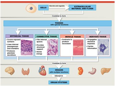
Four Major Types of Tissue
The human body contains four primary tissue types, each with distinct roles and characteristics:
Epithelial Tissue: Covers exposed surfaces, lines internal passageways, and forms glands.
Connective Tissue: Fills internal spaces, supports other tissues, transports materials, and stores energy.
Muscle Tissue: Specialized for contraction, enabling movement.
Nervous Tissue: Carries electrical signals throughout the body, facilitating communication.
Epithelial Tissue
Types and Functions
Epithelial tissue includes both epithelia and glands. Epithelia are layers of cells covering internal or external surfaces, while glands produce fluid secretions. The main functions of epithelial tissue are:
Physical Protection: Shields underlying tissues from mechanical and chemical injury.
Control Permeability: Regulates the movement of substances into and out of the body.
Provide Sensation: Contains sensory receptors for detecting environmental changes.
Produce Specialized Secretions: Forms glands that secrete substances for protection and communication.
Specializations and Characteristics
Epithelial cells exhibit several specializations and characteristics:
Polarity: Distinct apical (top) and basal (bottom) surfaces; apical surface may have microvilli or cilia.
Cellularity: Composed almost entirely of tightly packed cells with minimal extracellular matrix.
Attachment: Anchored to a basement membrane (basal lamina and reticular lamina).
Avascularity: Lacks blood vessels but is innervated.
Regeneration: Rapidly replaces lost cells by cell division.
Intercellular Connections
The integrity of epithelia is maintained by intercellular connections, attachment to the basement membrane, and epithelial maintenance and repair. Key cell junctions include:
Gap Junctions: Allow rapid communication and passage of ions and small molecules.
Tight Junctions: Prevent passage of water and solutes, maintaining compartmentalization.
Desmosomes: Provide strong adhesion, allowing tissues to withstand mechanical stress.
Classification of Epithelia
Shape and Layers
Epithelia are classified based on cell shape and the number of layers:
Shapes:
Squamous: Thin and flat.
Cuboidal: Square-shaped.
Columnar: Tall, slender rectangles.
Layers:
Simple: Single layer of cells.
Stratified: Several layers of cells.
Examples and Functions
Simple Squamous Epithelium: Absorption and diffusion; lines body cavities (mesothelium) and blood vessels (endothelium).
Stratified Squamous Epithelium: Protects against mechanical stresses; keratinized for strength and water resistance.
Simple Cuboidal Epithelium: Secretion and absorption; found in glands and kidney tubules.
Transitional Epithelium: Tolerates stretching; found in urinary bladder.
Simple Columnar Epithelium: Absorption and secretion; found in digestive tract.
Pseudostratified Columnar Epithelium: Typically ciliated; found in respiratory tract.
Connective Tissue
Components and Functions
Connective tissue consists of specialized cells, extracellular protein fibers, and ground substance. The matrix (fibers and ground substance) determines the tissue's function. Main functions include:
Establishing structural framework
Transporting fluids and dissolved materials
Protecting delicate organs
Supporting and interconnecting other tissues
Storing energy reserves
Defending against microorganisms
Categories of Connective Tissue
Connective Tissue Proper: Connects and protects (e.g., tendons, adipose tissue).
Fluid Connective Tissues: Transport (e.g., blood, lymph).
Supporting Connective Tissues: Structural strength (e.g., cartilage, bone).
Cells and Fibers of Connective Tissue Proper
Fibroblasts: Secrete proteins and hyaluronan for matrix formation.
Fibrocytes: Maintain connective tissue fibers.
Adipocytes: Store fat.
Mesenchymal Cells: Stem cells for repair.
Macrophages: Phagocytic immune cells.
Mast Cells: Release histamine and heparin for inflammation.
Lymphocytes: Immune response.
Microphages: Phagocytic blood cells.
Connective Tissue Fibers
Collagen Fibers: Strong, flexible, resist force in one direction.
Reticular Fibers: Network, resist forces in many directions.
Elastic Fibers: Stretchy, return to original length.
Loose and Dense Connective Tissues
Loose Connective Tissue: Areolar, adipose, and reticular tissues; fill spaces, cushion, and support.
Dense Connective Tissue: Regular, irregular, and elastic; provide strength and stability.
Fasciae
Superficial Fascia: Separates skin from underlying tissues.
Deep Fascia: Dense regular connective tissue sheets.
Subserous Fascia: Lies between deep fascia and serous membranes.
Fluid Connective Tissues: Blood and Lymph
Blood
Blood is a fluid connective tissue with a watery matrix (plasma) and formed elements:
Red Blood Cells (Erythrocytes): Transport oxygen.
White Blood Cells (Leukocytes): Immune defense.
Platelets (Thrombocytes): Blood clotting.
Lymph
Lymph forms as interstitial fluid enters lymphatic vessels, is monitored by the immune system, and returns to veins near the heart.
Supporting Connective Tissues: Cartilage and Bone
Cartilage
Cartilage provides shock absorption and protection. Its matrix is a firm gel containing chondroitin sulfates, and cells (chondrocytes) reside in lacunae. Types include:
Hyaline Cartilage: Tough, flexible, reduces friction; found in joints, ribs, sternum, trachea.
Elastic Cartilage: Supportive, bends easily; found in external ear, epiglottis.
Fibrocartilage: Durable, prevents bone-to-bone contact; found in joints, pubic bones, vertebrae.
Bone (Osseous Tissue)
Bones are rigid due to calcium salts and flexible collagen fibers. Osteocytes reside in lacunae, arranged around central canals. Periosteum covers bone, providing protection and growth.
Tissue Membranes
Types and Functions
Tissue membranes are physical barriers lining or covering body surfaces, consisting of epithelium supported by connective tissue. Four types:
Mucous Membranes: Line passageways with external connections; moist to reduce friction and facilitate absorption/secretion.
Serous Membranes: Line cavities not open to outside; parietal and visceral portions, serous fluid reduces friction.
Cutaneous Membrane: Skin; thick, waterproof, dry.
Synovial Membranes: Line joint cavities; produce synovial fluid for lubrication.
Muscle Tissue
Types and Features
Muscle tissue is specialized for contraction and includes three types:
Skeletal Muscle: Large, striated, voluntary; responsible for body movement.
Cardiac Muscle: Striated, involuntary; found only in the heart, regulated by pacemaker cells.
Smooth Muscle: Nonstriated, involuntary; found in walls of hollow organs, can divide and regenerate.
Nervous Tissue
Structure and Role
Nervous tissue conducts electrical impulses and is concentrated in the brain and spinal cord. It consists of:
Neurons: Nerve cells with cell body, dendrites (receive signals), and axon (transmits signals).
Neuroglia: Supporting cells that maintain and protect neurons.
Tissue Injuries and Repair
Response to Injury
Tissues respond to injury in two stages:
Inflammation: Triggered by trauma or infection; damaged cells release chemicals, mast cells activate.
Regeneration: Restores normal function; varies among tissues (epithelia and connective tissues regenerate well, muscle and nervous tissues poorly).
Aging and Tissue Structure
Effects of Aging
Aging decreases the speed and effectiveness of tissue regeneration due to hormonal changes and reduced activity. Effects include:
Thinner epithelia
Fragile connective tissues
Increased bruising
Brittle bones
Cardiovascular disease
Mental deterioration
Cancer rates increase with age, with environmental and chemical exposure as major causes.
