 Back
BackTissue Level of Organization: Structure and Function in Human Anatomy
Study Guide - Smart Notes
 Tailored notes based on your materials, expanded with key definitions, examples, and context.
Tissue Level of Organization
Definition and Overview
The tissue level of organization is a fundamental concept in anatomy and physiology, describing groups of cells with a common function and embryonic origin. Tissues are classified into four main types: epithelial, connective, muscle, and nervous tissue.
Epithelial tissue: Covers surfaces and secretes substances.
Connective tissue: Binds, supports, and protects other tissues.
Muscle tissue: Contracts to produce movement.
Nervous tissue: Communicates via action potentials.
Cell Junctions
Types and Functions
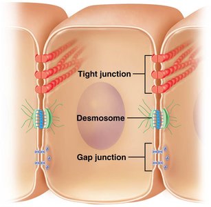
Cell junctions are points of contact between adjacent cell membranes, crucial for maintaining tissue integrity and communication.
Tight junctions: Prevent passage of substances between cells.
Desmosomes: Provide mechanical strength by anchoring cells together.
Hemidesmosomes: Attach cells to the basement membrane.
Adherens junctions: Connect actin filaments between cells.
Gap junctions: Allow direct communication via ions and small molecules.
Epithelial Tissue
Functions and Characteristics
Epithelial tissue serves as a selective barrier, regulates movement of materials, produces secretions, and protects surfaces from environmental damage.
Selective barrier: Controls entry and exit of substances.
Secretions: Lubrication, digestion, and protection.
Protection: Shields underlying tissues from physical and chemical harm.
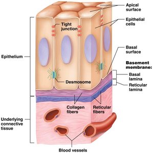
Structural Features
Has a nerve supply but lacks blood supply (avascular).
Cells attach to each other via cell junctions and to connective tissue via the basement membrane.
Covering epithelia: cells arranged in layers.
Secretory epithelia: cells often in clusters.
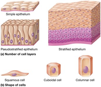
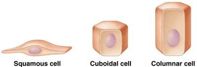
Epithelial Classification: Cell Shape
Epithelial cells are classified by shape:
Squamous: Flat cells.
Cuboidal: Equal width and height.
Columnar: Taller than wide.
Transitional: Variable shape, from squamous to cuboidal.
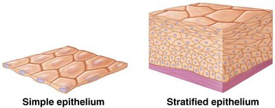
Epithelial Classification: Cell Layers
Classification by cell layers:
Simple: One layer.
Stratified: More than one layer; top layer determines category name.
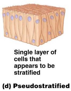
Pseudostratified: Appears layered, but all cells touch the basement membrane.
Covering and Lining Epithelia
Simple squamous: Diffusion and filtration (e.g., endothelium, mesothelium).
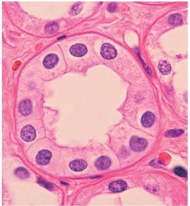
Simple cuboidal: Secretion and absorption (e.g., thyroid, kidneys).
Simple columnar: Non-ciliated for absorption; ciliated for moving fluids or particles.

Stratified squamous: Many layers, protective, high mitosis. Two types:
Non-keratinized (moist): Lines vaginal canal, GI tract.
Keratinized (dry): Skin, dead cells and keratin protein at surface.
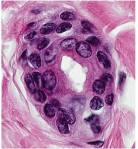
Stratified cuboidal: Uncommon, protection and secretion (e.g., sweat glands).
Stratified columnar: Rare, protection and secretion (e.g., salivary glands).
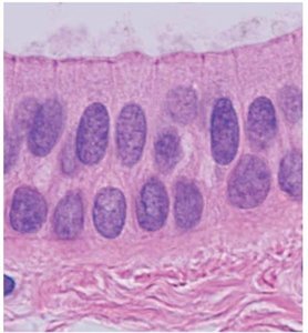
Pseudostratified: Often ciliated with goblet cells, lines respiratory tract.
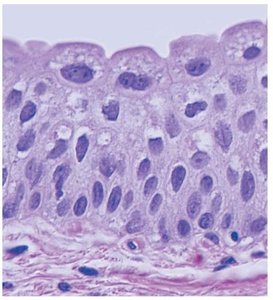
Transitional epithelium: Capable of stretching, uppermost cells are round, lines bladder, ureters, urethra.
Glandular Epithelia
Types and Functions
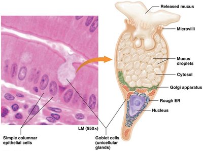
Unicellular: Release directly to surface (e.g., goblet cell).
Multicellular: Sweat, oil, salivary glands.
Endocrine glands: No ducts, secretions (hormones) released into blood.
Exocrine glands: Release secretions through ducts to surface.
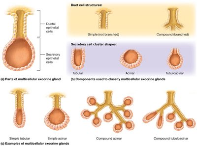
Exocrine Glands: Structural and Functional Classification
Structural: Duct portion (simple or compound), secretory portion (tubular, coiled, acinar).
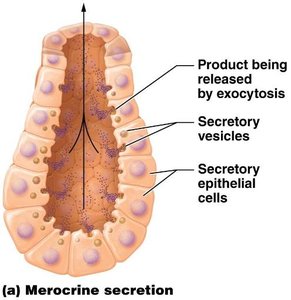
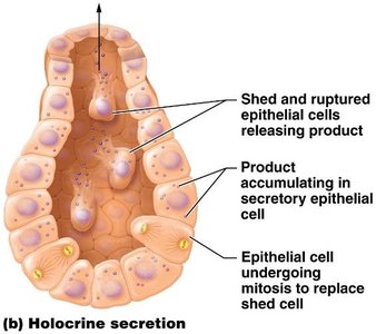
Functional:
Merocrine: Secretion only, released via vesicles (e.g., saliva).
Holocrine: Secretion released with entire cell (e.g., sebum).
Connective Tissue
General Structure and Functions
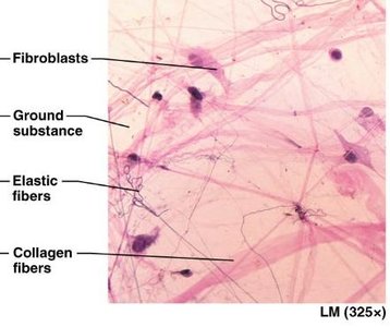
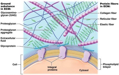
Connective tissue consists of cells and matrix (fibers and ground substance). It is vascular and innervated, providing support, protection, insulation, transport, immune responses, and energy storage.
Matrix: Fibers (collagen, elastin, reticular) and ground substance (various sulfates, hyaluronic acid).
Cells: Immature cells ("blast"), mature cells ("cyte"), mast, plasma, and blood cells.
Connective Tissue Subcategories
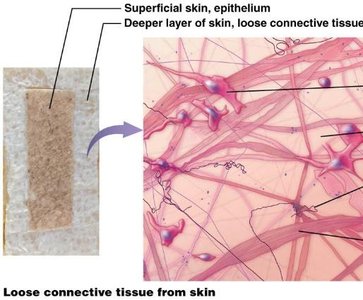
Areolar (loose): Common, support and flexibility, similar amounts of cells, fibers, and ground substance.
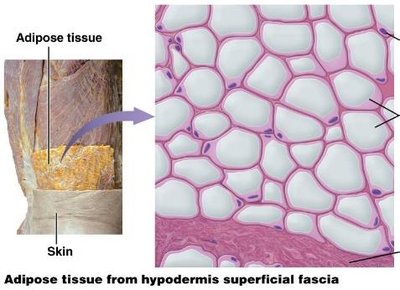
Adipose tissue: Common, insulation and energy reserves, white and brown types.
Reticular tissue: High reticular fibers, forms mesh, filters blood (spleen) or lymph (lymph node).
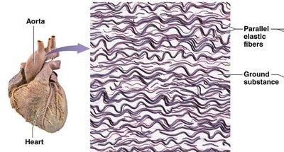
Elastic tissue: High elastic fibers and fibroblasts, in organs that must stretch and recoil.
Dense connective tissue: Fibers more abundant than cells or ground substance.
Dense irregular: No orientation to fibers, common in dermis, valves.
Dense regular: Parallel orientation to fibers, found in tendons and ligaments.
Cartilage: Chondrocytes in lacunae, three types:
Hyaline: Most common, collagen fibers and perichondrium.
Fibrocartilage: No perichondrium, strong, found in intervertebral discs.
Elastic: Elastic fibers and perichondrium, found in epiglottis, auricle of ear.
Bone: Ossified ground substance (Ca++, P), osteocytes in lacunae, two forms: compact and spongy.
Liquid connective tissues: Blood (cells, plasma) and lymph.
Muscle Tissue
Types and Functions
Muscle tissue contains contractile proteins (actin and myosin) and is classified into three types:
Skeletal muscle: Regular arrangement of proteins creates striations, voluntary, usually attached to bones.
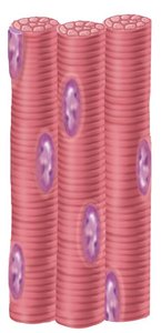
Cardiac muscle: Striations present, involuntary, intercalated discs connect cells for simultaneous contraction.
Smooth muscle: Involuntary, striations absent, found in walls of hollow structures.
Nervous Tissue
Cell Types and Functions
Nervous tissue is specialized for communication and control, consisting of two main cell types:
Neuroglia: Support function of neurons (six types).
Neurons: Generate and transmit electrical signals; cell extensions are critical to function.

Membranes
Types and Functions
Membranes are flat sheets of tissue that cover or line body parts. Epithelial membranes contain both epithelial and connective tissue.
Mucous membranes: Line body cavities opening to the exterior.
Serous membranes: Line body cavities that do not open to the exterior.
Cutaneous membrane: Covers the outside of the body; forms part of the integumentary system.
Synovial membrane: Lines most joint cavities, lacks epithelium, produces synovial fluid.
Tissue Repair
Capacity and Process
Tissue repair is the process of fixing or replacing worn out, damaged, or dead cells. Epithelial and connective tissues have a high capacity for repair due to increased mitotic ability, while muscle and nervous tissues have a lower capacity for repair.
Epithelial & connective tissues: High repair capacity.
Muscle & nervous tissues: Low repair capacity.
Mitotic ability: Directly correlates with repair capacity.
