 Back
BackTissue: The Living Fabric (Chapter 4) – ANP College Study Notes
Study Guide - Smart Notes
 Tailored notes based on your materials, expanded with key definitions, examples, and context.
Tissue: The Living Fabric
Introduction to Tissues
Tissues are groups of cells that are similar in structure and perform a common or related function. The human body is composed of four primary tissue types, each specialized for distinct roles essential to maintaining homeostasis.
Epithelial Tissue: Covers surfaces and lines cavities.
Connective Tissue: Supports, binds, and protects other tissues.
Muscle Tissue: Responsible for movement.
Nervous Tissue: Initiates and transmits electrical impulses.
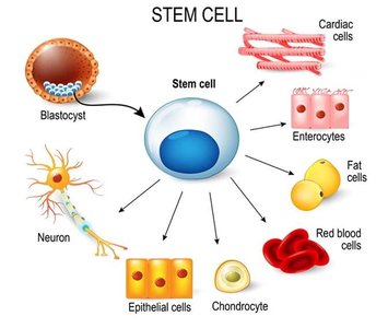
Epithelial Tissue
Structural and Functional Characteristics
Epithelial tissue (epithelium) forms boundaries between different environments, protects, secretes, absorbs, and filters. It covers body surfaces, lines internal cavities, and forms glands.
Apical Surface: The free surface exposed to the body exterior or cavity.
Basement Membrane: Anchors epithelium to underlying connective tissue.
Tightly Packed Cells: Provides a continuous layer for protection.
Avascular: Lacks blood vessels; nutrients diffuse from underlying tissues.
High Regenerative Capacity: Basal cells readily divide to replace lost cells.
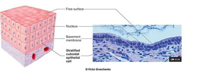
Classification of Epithelia
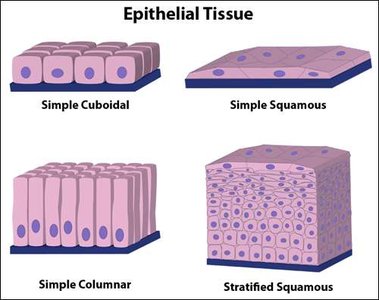
Epithelia are classified by the number of cell layers and the shape of cells at the apical surface.
Simple Epithelium: Single cell layer (absorption, secretion, filtration).
Stratified Epithelium: Multiple layers (protection).
Cell Shapes:
Squamous: Flat and scale-like.
Cuboidal: Box-like, as tall as they are wide.
Columnar: Tall and column-shaped.
Membranes Formed by Epithelia
Cutaneous Membrane: The skin; covers the body surface.
Mucous Membranes (Mucosae): Line body cavities open to the exterior (e.g., digestive, respiratory tracts).
Serous Membranes (Serosae): Line closed ventral body cavities; secrete serous fluid to reduce friction.
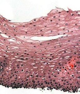
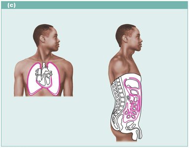
Glands and Glandular Epithelium
A gland consists of one or more cells that make and secrete a particular product (secretion). Glands are classified by where they release their product and the number of cells they contain.
Endocrine Glands: Ductless; secrete hormones directly into blood or tissue fluids.
Exocrine Glands: Secrete products into ducts that open onto body surfaces (e.g., sweat, salivary glands).
Unicellular Glands: Single-celled (e.g., goblet cells).
Multicellular Glands: Composed of many cells; classified by duct structure and secretory units.
Structural Classification of Multicellular Exocrine Glands
Simple Glands: Unbranched duct.
Compound Glands: Branched duct.
Tubular: Secretory cells form tubes.
Alveolar (Acinar): Secretory cells form sacs.
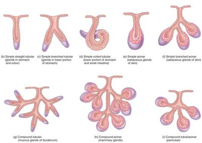
Modes of Secretion
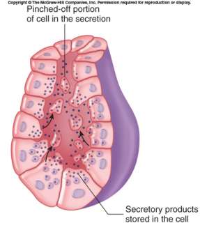
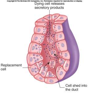
Merocrine: Secrete by exocytosis (e.g., sweat glands).
Apocrine: Pinch off portions of the cell (e.g., mammary glands).
Holocrine: Entire cell ruptures to release product (e.g., sebaceous glands).
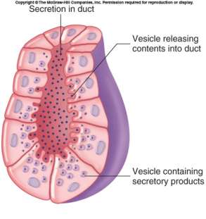
Connective Tissue
General Characteristics
Connective tissue is the most abundant and widely distributed tissue type. It supports, protects, and binds other tissues together. All connective tissues share three basic components: cells, fibers, and ground substance (together forming the extracellular matrix).
Cells: Vary by tissue type (e.g., fibroblasts, chondrocytes, osteocytes, adipocytes).
Matrix: Nonliving material between cells; consists of fibers and ground substance.
Connective Tissue Fibers
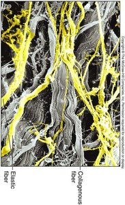
Collagen Fibers: Strong, provide tensile strength ("white fibers").
Elastic Fibers: Stretch and recoil ("yellow fibers").
Reticular Fibers: Short, fine, branched; support soft tissues.
Ground Substance
The ground substance fills the space between cells and fibers. It consists of:
Hyaluronic Acid: Lubricant.
Proteoglycans: Trap water, affecting tissue viscosity.
Adhesive Molecules: Glue cells to matrix elements.
Classification of Connective Tissue
Connective Tissue Proper:
Loose Connective Tissue: Areolar, adipose, reticular.
Dense Connective Tissue: Dense regular, dense irregular, elastic.
Specialized Connective Tissue: Cartilage, bone, blood.
Areolar Tissue (Loose Connective Tissue)
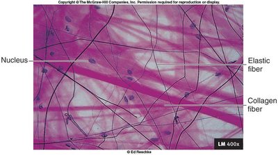
Cells: Fibroblasts, macrophages, mast cells, some white blood cells.
Fibers: Collagen, elastic, reticular.
Function: Wraps and cushions organs; holds tissue fluid.
Location: Widely distributed under epithelia, surrounds capillaries.
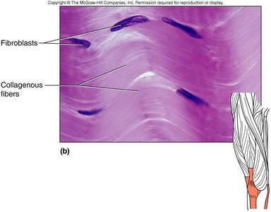
Dense Connective Tissue
Cells: Mainly fibroblasts.
Fibers: Densely packed collagen fibers.
Function: Attaches muscles to bones or to muscles; withstands tensile stress.
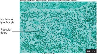
Reticular Connective Tissue
Cells: Reticular cells, lymphocytes.
Fibers: Reticular fibers.
Function: Forms a soft internal skeleton (stroma) that supports other cell types.
Location: Lymphoid organs (lymph nodes, bone marrow, spleen).
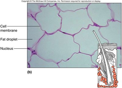
Adipose Tissue (Fat)
Cells: Adipocytes.
Fibers: Few collagenous fibers.
Function: Provides cushioning, insulation, and energy storage.
Location: Under skin, around vital organs, behind eyes.
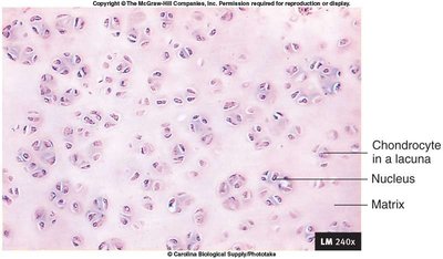
Cartilage
Cells: Chondrocytes in lacunae.
Matrix: Contains collagen or elastic fibers and ground substance.
Types:
Hyaline Cartilage: Most common; supports and reinforces.
Elastic Cartilage: Maintains shape and flexibility.
Fibrocartilage: Absorbs compressive shock; found in intervertebral discs.
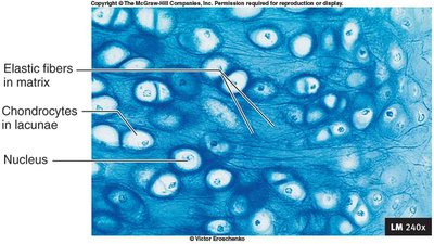
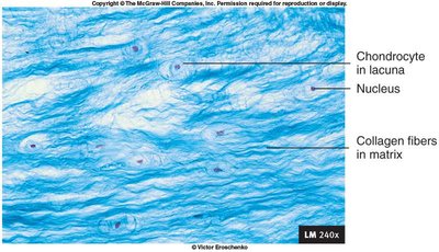
Bone and Blood
Bone:
Cells: Osteocytes in lacunae.
Matrix: Hard, calcified with collagen fibers.
Function: Supports, protects, stores minerals.
Blood:
Cells: Red blood cells, white blood cells, platelets.
Matrix: Plasma (fluid).
Function: Transport of respiratory gases, nutrients, wastes.
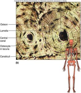
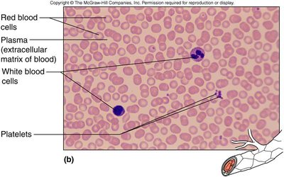
Additional info: This guide covers the essential structural and functional features of epithelial and connective tissues, including their classification, cellular components, and representative images for histological identification. These concepts are foundational for understanding tissue organization in human anatomy and physiology.
