 Back
Tissue: The Living Fabric – Connective, Muscle, and Nervous Tissues
Study Guide - Smart Notes
 Tailored notes based on your materials, expanded with key definitions, examples, and context.
Connective Tissue
Overview of Connective Tissue
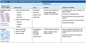
Connective tissue is the most abundant and widely distributed of the primary tissue types in the human body. Its major functions include binding and supporting other tissues, protecting organs, insulating the body, storing reserve fuel, and transporting substances such as blood.
Four main classes: Connective tissue proper, cartilage, bone, and blood.
Key functions: Support, protection, insulation, energy storage, and transport.
Common Characteristics of Connective Tissue
Embryonic origin: All connective tissues arise from mesenchyme tissue.
Vascularity: Varies from avascular (cartilage) to highly vascularized (bone).
Extracellular matrix (ECM): Cells are suspended in a protein-sugar mesh that supports and protects them.
Structural Elements of Connective Tissue
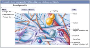
All connective tissues share three main structural elements:
Ground substance: Unstructured gel-like material that fills the space between cells and allows diffusion of nutrients and waste.
Fibers: Provide support and include collagen (strength), elastic (stretch and recoil), and reticular (supportive networks).
Cells: "Blast" cells (immature, matrix-secreting) and "cyte" cells (mature, maintenance).
Types of Connective Tissue Proper
Loose connective tissues: Areolar, adipose, and reticular.
Dense connective tissues: Dense regular, dense irregular, and elastic.
Loose Connective Tissues
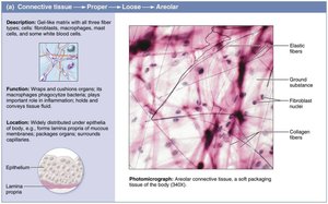
Areolar: Most widely distributed, supports and binds tissues, holds body fluids, defends against infection, stores nutrients. Contains fibroblasts, macrophages, and fat cells.
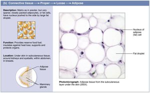
Adipose: Stores energy, insulates, and cushions organs. Adipocytes are the main cell type.
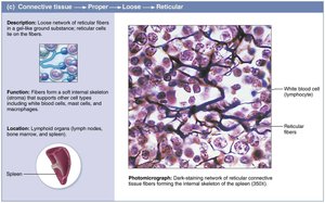
Reticular: Forms a soft internal skeleton (stroma) that supports other cell types in lymphoid organs.
Dense Connective Tissues
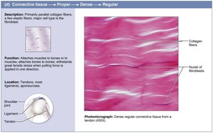
Dense regular: Parallel collagen fibers, great tensile strength, found in tendons and ligaments.
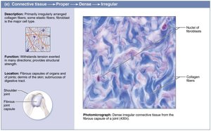
Dense irregular: Irregularly arranged collagen fibers, withstands tension from multiple directions, found in dermis and joint capsules.
Elastic: High proportion of elastic fibers, allows tissue to recoil after stretching, found in large arteries.
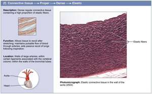
Cartilage
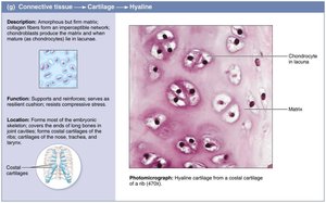
Cartilage is a tough yet flexible tissue that lacks nerve fibers and is avascular. It is composed of chondroblasts (during growth) and chondrocytes (in lacunae), with a matrix that is 80% water.
Hyaline cartilage: Most abundant, supports and reinforces, found at ends of long bones, nose, trachea, and ribs.
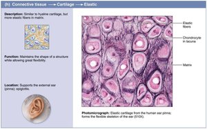
Elastic cartilage: More elastic fibers, maintains shape of structures like the ear and epiglottis.
Fibrocartilage: Thick collagen fibers, strong and resists compression, found in intervertebral discs and knee.
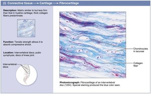
Bone (Osseous Tissue)
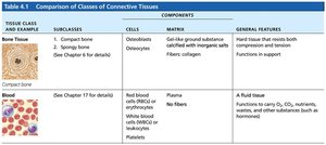
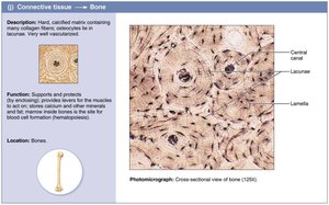
Bone tissue supports and protects body structures, stores fat, and synthesizes blood cells. It is highly vascularized and contains more collagen than cartilage, as well as inorganic calcium salts. Osteoblasts produce the matrix, and osteocytes maintain it.
Blood
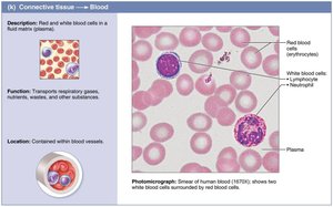
Blood is a fluid connective tissue consisting of cells (red and white blood cells, platelets) suspended in plasma. It functions in the transport of respiratory gases, nutrients, wastes, and other substances throughout the body.
Muscle Tissue
Overview of Muscle Tissue
Muscle tissue is highly vascularized and responsible for most types of body movement. Muscle cells contain myofilaments made of actin and myosin, which enable contraction. There are three types of muscle tissue: skeletal, cardiac, and smooth.
Skeletal Muscle
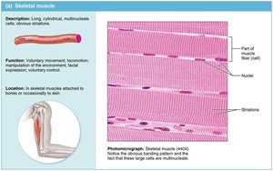
Structure: Long, cylindrical, multinucleate cells with obvious striations.
Function: Voluntary movement, locomotion, facial expression.
Location: Attached to bones or occasionally to skin.
Cardiac Muscle
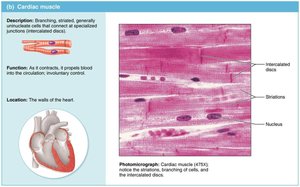
Structure: Branching, striated, generally uninucleate cells connected at intercalated discs.
Function: Involuntary control; propels blood into circulation.
Location: Walls of the heart.
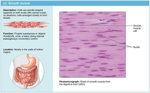
Smooth Muscle
Structure: Spindle-shaped cells with central nuclei, no striations, form sheets.
Function: Involuntary control; propels substances or objects along internal passageways.
Location: Walls of hollow organs (except the heart).
Nervous Tissue
Overview of Nervous Tissue
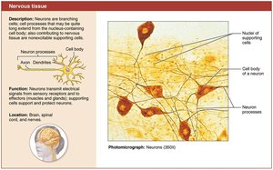
Nervous tissue is the main component of the nervous system, which includes the brain, spinal cord, and nerves. It regulates and controls body functions through the generation and conduction of nerve impulses.
Neurons: Specialized cells that generate and conduct electrical impulses.
Supporting cells: Support, insulate, and protect neurons.
Covering and Lining Membranes
Types of Membranes
Membranes are composed of at least two primary tissue types: an epithelium bound to underlying connective tissue. The three main types are cutaneous, mucous, and serous membranes.
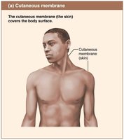
Cutaneous membrane: The skin; a dry membrane covering the body surface.
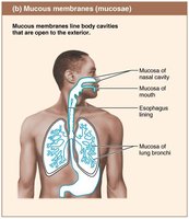
Mucous membranes: Line body cavities open to the exterior (digestive, respiratory, urogenital tracts); moist and may secrete mucus.
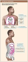
Serous membranes: Line closed ventral body cavities; consist of mesothelium and areolar connective tissue. Parietal serosae line cavity walls, visceral serosae cover organs. Examples: pleurae (lungs), pericardium (heart), peritoneum (abdomen).
Tissue Repair
Steps in Tissue Repair
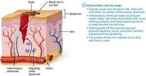
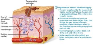
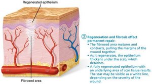
Tissue repair occurs in three main steps: inflammation, organization, and regeneration/fibrosis.
Inflammation: Inflammatory chemicals are released, blood vessels dilate, and clotting occurs to prevent infection and further injury.
Organization: The blood clot is replaced by granulation tissue, fibroblasts produce collagen, and macrophages remove debris.
Regeneration and fibrosis: The epithelium regenerates, the scab detaches, and scar tissue may form.
Regenerative Capacity of Different Tissues
High regenerative capacity: Epithelial tissues, bone, areolar connective tissue, dense irregular connective tissue, blood-forming tissue.
Moderate capacity: Smooth muscle, dense regular connective tissue.
Low to none: Cardiac muscle, nervous tissue in brain and spinal cord.
Developmental Aspects of Tissues
Primary Germ Layers
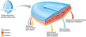
During embryonic development, three primary germ layers form: ectoderm, mesoderm, and endoderm. These layers give rise to all tissues in the body.
Ectoderm: Forms nerve tissue.
Mesoderm: Forms muscle and connective tissues.
Endoderm: Contributes to epithelial linings of internal organs.
Additional info: As the body ages, tissue repair becomes less efficient, and tissues such as bone, muscle, and nervous tissue may atrophy. DNA mutations increase the risk of cancer.
