 Back
BackTissue 1: The Living Fabric – Epithelial Tissue Study Guide
Study Guide - Smart Notes
 Tailored notes based on your materials, expanded with key definitions, examples, and context.
Tissue: The Living Fabric
Introduction to Tissues
Tissues are groups of cells with similar structure and function, working together to maintain homeostasis in the human body. The study of tissues is known as histology.
Definition: Tissue – a collection of cells performing a common or related function.
Importance: Specialized cells in tissues ensure proper functioning of organs and systems.
Types of Primary Tissues
The human body contains four primary tissue types, each with distinct roles:
Epithelial tissue: Covers surfaces, forms boundaries, protects, secretes, absorbs, and filters.
Connective tissue: Supports, protects, and binds other tissues.
Muscle tissue: Produces movement by contraction.
Nervous tissue: Controls and communicates via electrical signals.
Epithelial Tissue
Overview and Functions
Epithelial tissue forms boundaries between different environments and is found on external and internal surfaces, as well as in glands. It serves several essential functions:
Protection: Shields underlying tissues from injury and infection.
Absorption: Takes in substances such as nutrients.
Filtration: Selectively allows passage of materials.
Excretion: Removes waste products.
Secretion: Produces and releases substances.
Sensory reception: Detects changes in the environment.
Characteristics of Epithelial Tissue
Epithelial tissues possess five key characteristics:
Polarity: Cells have distinct apical (upper) and basal (lower) surfaces.
Specialized contacts: Cells are tightly joined by junctions (tight junctions, desmosomes).
Supported by connective tissues: The basement membrane (basal lamina + reticular lamina) reinforces the tissue.
Avascular but innervated: No blood vessels; nutrients diffuse from underlying tissues. Supplied by nerve fibers.
Regeneration: High capacity for renewal, especially when exposed to friction or hostile environments.
Polarity in Epithelial Tissue
The apical surface may be smooth, have microvilli (increasing surface area, e.g., intestinal lining), or cilia (e.g., trachea). The basal surface is attached to a noncellular basal lamina, which acts as an adhesive sheet, selective filter, and scaffolding for cell migration during repair.
Specialized Contacts
Epithelial cells form continuous sheets, bound by lateral contacts such as tight junctions and desmosomes, ensuring integrity and selective permeability.
Connective Tissue Support
All epithelial tissues are supported by connective tissue, with the basement membrane providing structural reinforcement and defining boundaries.
Avascular but Innervated
Epithelial tissues lack blood vessels and rely on diffusion for nourishment, but are richly supplied with nerves for sensory functions.
Regeneration
Epithelial tissues can rapidly replace lost cells through cell division, especially when exposed to friction or harmful substances, provided adequate nutrients are available.
Classification of Epithelia
Layer and Shape
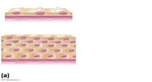
All epithelial tissues are classified by two criteria: number of cell layers and cell shape.
Simple epithelia: Single layer of cells.
Stratified epithelia: Two or more layers; named by the shape of cells in the apical layer.
Cell shapes:
Squamous: Flattened, scalelike; nucleus flattened.
Cuboidal: Boxlike; nucleus round.
Columnar: Tall, column-shaped; nucleus elongated.
Example: Simple squamous epithelium lines blood vessels; stratified squamous epithelium forms the skin surface.
Glandular Epithelia
Glands and Secretion
A gland consists of one or more cells that produce and secrete an aqueous fluid called a secretion. Glands are classified by site of product release (endocrine or exocrine) and number of cells (unicellular or multicellular).
Endocrine glands: Ductless; secrete hormones into blood or lymph, affecting target organs.
Exocrine glands: Secrete products into ducts, releasing onto body surfaces or cavities (e.g., sweat, oil, salivary glands).
Unicellular Exocrine Glands
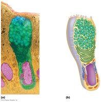
The most important unicellular exocrine glands are mucous cells and goblet cells, found in epithelial linings of the intestinal and respiratory tracts. They produce mucin, which dissolves in water to form mucus—a protective, lubricating coating.
Multicellular Exocrine Glands
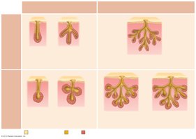
Multicellular exocrine glands consist of a duct and a secretory unit, often surrounded by supportive connective tissue that supplies blood and nerve fibers. These glands are classified by structure and type of secretion:
Structure: Simple (unbranched duct) or compound (branched duct); cells may be tubular, alveolar, or tubuloalveolar.
Type of secretion:
Merocrine: Secrete products by exocytosis as produced (e.g., pancreas, salivary glands, most sweat glands).
Holocrine: Accumulate products within, then rupture (e.g., sebaceous glands).
Apocrine: Accumulate products, only apex ruptures (controversial in humans).
Modes of Secretion in Exocrine Glands
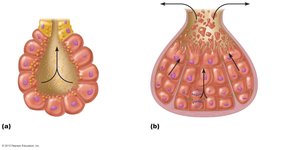
Exocrine glands release their products by different mechanisms:
Merocrine: Products are secreted by exocytosis.
Holocrine: Entire secretory cell ruptures, releasing secretions and cell fragments.
Summary Table: Classification of Epithelial Tissue
Layer | Shape | Example |
|---|---|---|
Simple | Squamous | Blood vessel lining |
Simple | Cuboidal | Kidney tubules |
Simple | Columnar | Intestinal lining |
Stratified | Squamous | Skin surface |
Stratified | Cuboidal | Rare, sweat glands |
Stratified | Columnar | Rare, male urethra |
Additional info: The notes above expand on the original slides by providing definitions, examples, and context for each tissue type and gland classification, ensuring completeness and academic quality for exam preparation.
