 Back
BackTissue: The Living Fabric – Structure, Function, and Classification
Study Guide - Smart Notes
 Tailored notes based on your materials, expanded with key definitions, examples, and context.
Introduction to Tissues and Histology
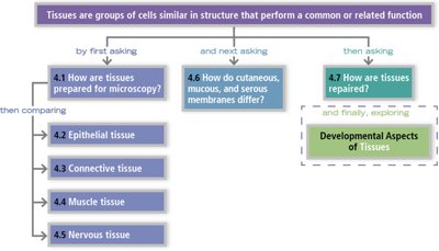
Tissues are groups of cells similar in structure that perform a common or related function. The study of tissues, known as histology, is fundamental to understanding the organization and function of the human body. Histological analysis involves preparing tissue samples, staining them, and examining them under various types of microscopes to reveal their structure and organization.
Preparation and Study of Tissues
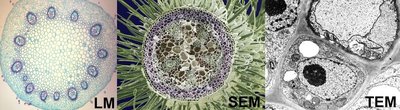
Microscopy and Staining
Fixation: Preserves tissue structure using chemicals like formalin or by freezing.
Sectioning: Thin slices are cut for microscopic examination.
Staining: Dyes are used to enhance contrast and highlight specific structures.
Microscopy Types:
Light Microscopy (LM): Views stained tissues in color.
Transmission Electron Microscopy (TEM): Provides high-magnification grayscale images of internal structures.
Scanning Electron Microscopy (SEM): Offers 3D views of tissue surfaces.
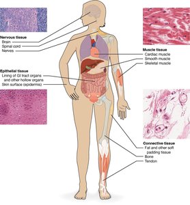
Major Tissue Types in the Human Body
The human body is composed of four primary tissue types, each with distinct structures and functions:
Epithelial Tissue
Connective Tissue
Muscle Tissue
Nervous Tissue
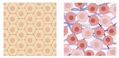
Epithelial Tissue
Characteristics and Functions
Polarity: Has an apical (exposed) and basal (attached) surface.
Specialized Contacts: Tight junctions and desmosomes bind cells together.
Regeneration: High capacity for renewal.
Avascular but Innervated: Lacks blood vessels but contains nerves.
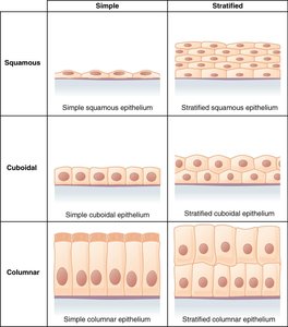
Classification of Epithelia
Epithelia are classified by the number of layers and the shape of cells:
Simple | Stratified | |
|---|---|---|
Squamous | Simple squamous epithelium | Stratified squamous epithelium |
Cuboidal | Simple cuboidal epithelium | Stratified cuboidal epithelium |
Columnar | Simple columnar epithelium | Stratified columnar epithelium |
Special Epithelia
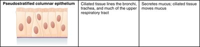
Pseudostratified columnar epithelium | Description | Function |
|---|---|---|
| Ciliated tissue lines the bronchi, trachea, and much of the upper respiratory tract | Secretes mucus; ciliated tissue moves mucus |
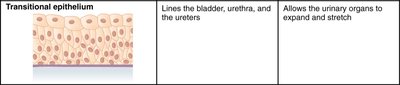
Transitional epithelium | Description | Function |
|---|---|---|
| Lines the bladder, urethra, and the ureters | Allows the urinary organs to expand and stretch |
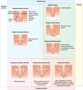
Glandular Epithelium
Glands: One or more cells that produce and secrete a specific product.
Endocrine Glands: Ductless; secrete hormones directly into tissues (e.g., thyroid hormone, insulin).
Exocrine Glands: Secrete products into ducts leading to epithelial surfaces (e.g., sweat, saliva).
Unicellular Glands: Goblet cells.
Multicellular Glands: Can be tubular or alveolar (acinar) in structure.
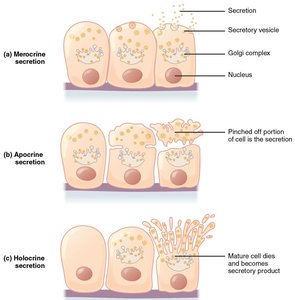
Modes of Secretion
Merocrine: Secretion by exocytosis (e.g., sweat glands).
Apocrine: Part of the cell pinches off (e.g., mammary glands).
Holocrine: Entire cell ruptures, releasing product (e.g., sebaceous glands).
Connective Tissue
General Features
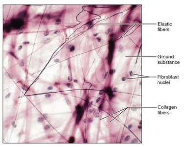
Composed of loosely dispersed cells in an extensive extracellular matrix.
Ground Substance: Fills space between cells; composed of interstitial fluid, proteins, and polysaccharides.
Protein Fibers: Collagen (strength), elastic (stretch), and reticular (support).
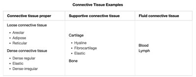
Classification of Connective Tissue
Connective tissue proper | Supportive connective tissue | Fluid connective tissue |
|---|---|---|
Loose: Areolar, Adipose, Reticular Dense: Dense regular, Elastic, Dense-irregular | Cartilage: Hyaline, Fibrocartilage, Elastic Bone | Blood Lymph |
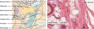
Cells of Connective Tissue Proper
Fibroblasts/Fibrocytes: Produce fibers and ground substance.
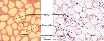
Adipocytes: Store fat (white and brown types).
Mesenchymal Stem Cells: Differentiate into other cell types.
Macrophages: Phagocytize debris and pathogens.
Mast Cells: Release histamine in inflammation.
Loose Connective Tissue
Areolar: Most common; supports and binds other tissues.
Adipose: Stores energy, insulates, and cushions organs.
Reticular: Forms a soft internal skeleton for lymphoid organs.
Dense Connective Tissue
Dense Regular: Parallel collagen fibers; found in tendons and ligaments.
Dense Irregular: Irregularly arranged fibers; found in dermis.
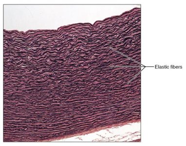
Elastic: High proportion of elastic fibers; found in large arteries.
Supportive Connective Tissue
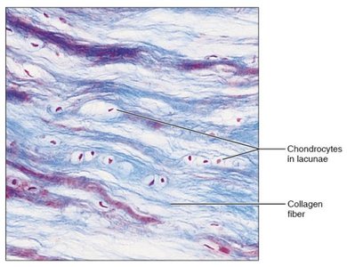
Cartilage: Chondrocytes in lacunae, avascular, flexible support.
Types: Hyaline, fibrocartilage, elastic cartilage.
Bone: Collagen fibers in a mineralized matrix (hydroxyapatite); highly vascularized.
Fluid Connective Tissue
Blood: Transports nutrients, gases, wastes, and cells.
Lymph: Returns fluid to blood, involved in immune responses.
Muscle Tissue
Muscle tissue is specialized for contraction and movement. There are three types:
Skeletal Muscle: Voluntary, striated, attached to bones.
Cardiac Muscle: Involuntary, striated, found in the heart.
Smooth Muscle: Involuntary, non-striated, found in walls of hollow organs.
Nervous Tissue
Nervous tissue is specialized for communication via electrical and chemical signals.
Neurons: Transmit electrical impulses (action potentials).
Neuroglia: Support, protect, and nourish neurons.
Key neuroglia: Astrocytes (CNS homeostasis), oligodendrocytes (CNS myelin), Schwann cells (PNS myelin).
Body Membranes
Cutaneous Membrane: Skin; dry, keratinized epithelium attached to connective tissue.
Mucous Membranes (Mucosae): Line body cavities open to the exterior; moist, adapted for absorption and secretion.
Serous Membranes (Serosae): Line closed ventral body cavities; consist of simple squamous epithelium over areolar connective tissue, secrete serous fluid.
Tissue Repair
Tissue repair involves restoring tissue integrity after injury through two main processes:
Regeneration: Replacement of destroyed tissue with the same tissue type.
Fibrosis: Replacement with scar tissue (dense connective tissue).
Inflammation: White blood cells, fluid, and proteins enter the area; clot forms.
Organization: New capillaries grow, collagen is produced, debris is cleared.
Permanent Repair: New tissue matures and restores function.
Summary Table: Tissue Types and Functions
Tissue Type | Main Function | Location Example |
|---|---|---|
Epithelial | Protection, absorption, secretion | Skin, lining of GI tract |
Connective | Support, binding, transport | Tendons, bone, blood |
Muscle | Movement | Skeletal muscles, heart |
Nervous | Communication, control | Brain, nerves |
