 Back
Tissues and Histology: Structure and Function of Epithelial, Connective, Muscle, and Nervous Tissues
Study Guide - Smart Notes
 Tailored notes based on your materials, expanded with key definitions, examples, and context.
Tissues & Histology
Overview of Tissues
Tissues are groups of cells with similar structure and function, forming the basic building blocks of organs and the body. The four primary tissue types are epithelial, connective, muscle, and nervous tissues. Each type has specialized roles essential for maintaining homeostasis and supporting life.
Epithelial Tissue
Classification of Epithelia
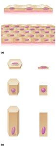
Epithelial tissues cover body surfaces, line cavities, and form glands. They are classified based on the number of cell layers and the shape of the cells at the apical surface.
Simple epithelium: Single layer of cells; specialized for absorption, secretion, and filtration.
Stratified epithelium: Multiple layers; provides protection in areas of high abrasion.
Cell shapes: Squamous (flat), Cuboidal (cube-shaped), Columnar (tall and column-like).
Formation of Glands
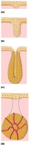
Glands are formed by the invagination of epithelial sheets. They are classified as exocrine (secrete into ducts) or endocrine (secrete into blood).
Exocrine glands: Maintain ducts to the epithelial surface (e.g., sweat, salivary glands).
Endocrine glands: Lose their ducts during development and release hormones directly into the bloodstream.
Types of Epithelial Tissues
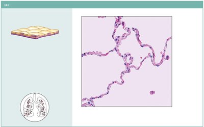
Simple squamous epithelium: Single layer of flat cells; allows diffusion and filtration. Location: Air sacs of lungs, lining of blood vessels.
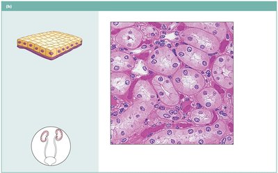
Simple cuboidal epithelium: Single layer of cube-shaped cells; functions in secretion and absorption. Location: Kidney tubules, small glands.
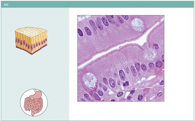
Simple columnar epithelium: Single layer of tall cells; absorption and secretion, may have cilia or goblet cells. Location: Digestive tract, uterine tubes.
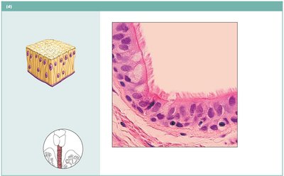
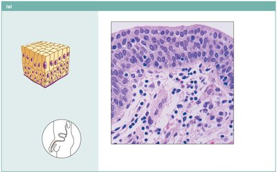
Pseudostratified columnar epithelium: Appears stratified but is a single layer; often ciliated with goblet cells. Location: Trachea, upper respiratory tract.
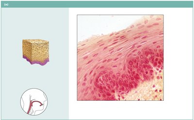
Stratified squamous epithelium: Multiple layers; protects against abrasion. Location: Skin, mouth, esophagus.
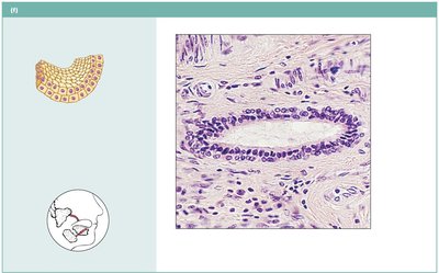
Stratified cuboidal/columnar epithelium: Rare; found in ducts of large glands and male urethra.
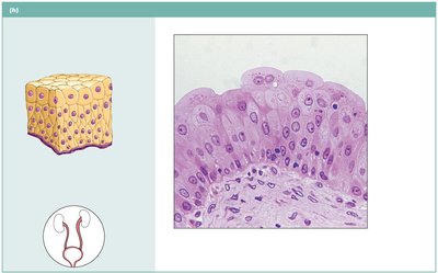
Transitional epithelium: Resembles both stratified squamous and cuboidal; stretches to accommodate urine. Location: Urinary bladder.
Connective Tissue
General Structure and Function
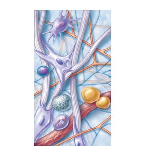
Connective tissues support, bind, and protect other tissues and organs. They are characterized by cells scattered within an extracellular matrix composed of protein fibers and ground substance.
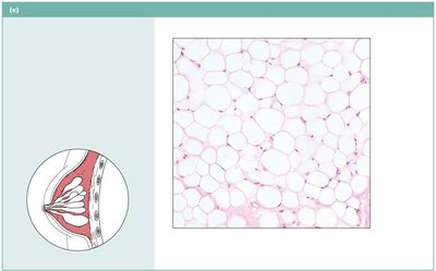
Major cell types: Fibroblasts, macrophages, mast cells, adipocytes, and white blood cells.
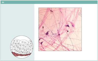
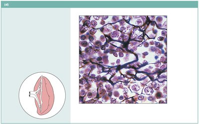
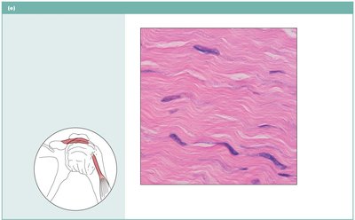
Fibers: Collagen (strength), elastic (flexibility), reticular (support).
Types of Connective Tissue
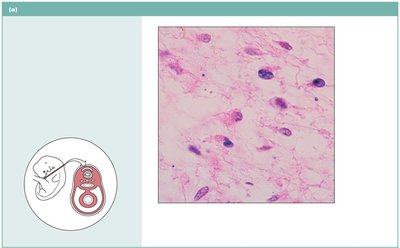
Loose connective tissue: Includes areolar, adipose, and reticular tissues. Functions in support, storage, and immunity.
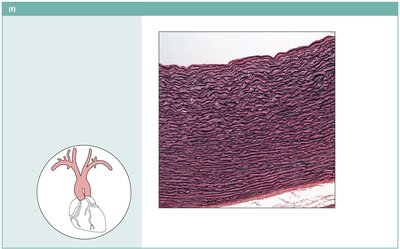
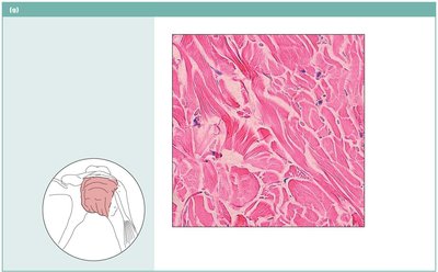
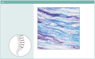
Dense connective tissue: Includes dense regular, dense irregular, and elastic tissues. Provides strength and elasticity.
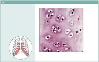
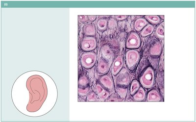
Cartilage: Hyaline, elastic, and fibrocartilage; provides support and flexibility.
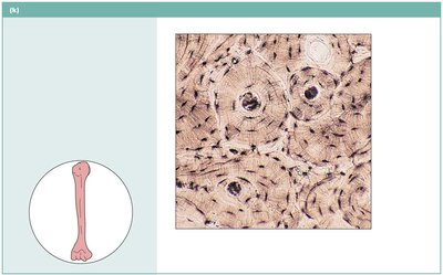
Bone (osseous tissue): Supports and protects; site of blood cell formation.
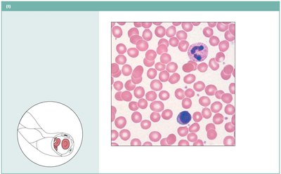
Blood: Transports gases, nutrients, and wastes.
Nervous Tissue
Structure and Function
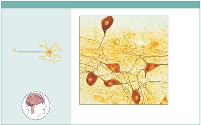
Nervous tissue is specialized for communication via electrical impulses. It consists of neurons and supporting cells (neuroglia).
Neurons: Large, branching cells that transmit electrical signals.
Supporting cells: Protect, insulate, and support neurons.
Location: Brain, spinal cord, nerves.
Muscle Tissue
Types of Muscle Tissue
Muscle tissue is responsible for movement and force generation. There are three types:
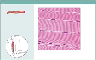
Skeletal muscle: Long, multinucleate, striated cells; voluntary movement. Location: Attached to bones.
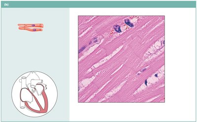
Cardiac muscle: Branching, striated, usually uninucleate cells with intercalated discs; involuntary. Location: Heart walls.
Smooth muscle: Spindle-shaped, non-striated cells; involuntary. Location: Walls of hollow organs.
Summary Table: Major Tissue Types and Functions
Tissue Type | Main Function | Key Locations |
|---|---|---|
Epithelial | Protection, absorption, secretion, filtration | Body surfaces, lining of organs, glands |
Connective | Support, binding, protection, transport | Bones, tendons, fat, blood, cartilage |
Muscle | Movement, force generation | Skeletal muscles, heart, walls of hollow organs |
Nervous | Communication, control | Brain, spinal cord, nerves |
Additional info: This summary integrates textbook-style explanations, definitions, and examples for each tissue type, with images included only where they directly reinforce the content.
