 Back
BackTissues and the Integumentary System: Structure, Function, and Repair
Study Guide - Smart Notes
 Tailored notes based on your materials, expanded with key definitions, examples, and context.
Tissue Level of Organization
Cell Junctions
Cell junctions are specialized structures that connect adjacent cells, providing structural integrity and facilitating communication.
Tight Junctions: Form a seal between cells, preventing the free passage of molecules (including ions) between epithelial cells.
Desmosomes: Provide strong adhesion between cells using cytoskeletal connections; found in tissues experiencing mechanical stress (e.g., cardiac muscle, bladder, gastrointestinal mucosa, epithelia).
Gap Junctions: Allow exchange of ions, secondary messengers, and small metabolites between adjacent cells; present in connective, epithelial, neural, and muscular tissues. Intercalated discs in cardiac muscle contain both desmosomes and gap junctions.
Major Tissue Types
Epithelial Tissue: Lines cavities and covers surfaces of blood vessels and organs; composed of closely packed, similar cells; avascular; good regeneration; located above the basement membrane.
Connective Tissue: Supports, connects, and separates different body tissues; varied cell types scattered in an extracellular matrix; contains blood capillaries (with some exceptions); regeneration varies; located below the basement membrane.
Muscle Tissue: Responsible for movement.
Nervous Tissue: Conducts electrical impulses and supports neural function.
Epithelial Tissue Classification
Epithelial tissues are classified by the number of layers and cell shape:
Layers: Simple (single layer), Stratified (multiple layers)
Shapes: Squamous (flat), Cuboidal (cube-shaped), Columnar (tall)
Special Types: Transitional (stratified, rounded cells that flatten), Pseudostratified (appears layered due to varying cell heights)
Examples: Simple squamous (one flat layer), Stratified columnar (multiple layers, taller than wide)
Basement Membrane
The basement membrane separates and protects tissues from mechanical stress. It consists of:
Basal Lamina: Extracellular matrix beneath epithelial/endothelial cells, muscle, and fat cells.
Reticular Lamina: Composed of collagen and reticulin fibers, providing support.
Functions of Epithelial Tissue Types
Simple Squamous: Lines blood vessels, capillaries, alveoli; functions in diffusion, filtration, osmosis.
Stratified Squamous: Found in skin, cheeks, vagina, esophagus; protects from abrasion; may contain keratin and melanin.
Pseudostratified: Lines trachea, upper respiratory tract, sperm ducts; functions in secretion and propulsion of mucus.
Transitional: Found in ureters, bladder, part of urethra; stretches to permit distension.
Simple Columnar: Lines intestines, stomach; functions in absorption and secretion.
Glands and Modes of Secretion
Glands are composed of epithelial tissue and classified as:
Endocrine: Secrete into the bloodstream.
Exocrine: Secrete into ducts onto organ surfaces.
Unicellular Glands: Goblet cells secrete mucus.
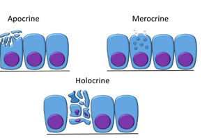
Modes of Secretion:
Merocrine: Secretion by exocytosis (e.g., sweat, salivary glands).
Apocrine: Secretion involves loss of part of the cell (e.g., mammary glands).
Holocrine: Entire cell disintegrates to release secretion (e.g., sebaceous glands).
Connective Tissue Overview
Connective tissue originates from mesenchyme (mesoderm) and consists of cells and extracellular matrix.
Cells: Fibroblasts (form fibers), adipocytes (store lipids), chondrocytes (maintain cartilage), osteocytes (maintain bone), osteoblasts (build bone), RBCs, WBCs, macrophages, mast cells.
Matrix: Ground substance (e.g., hyaluronic acid, calcium salts, chondroitin sulfate, glucosamine, plasma) and fibers (collagen, elastic, reticular).
Fiber Types:
Collagen: Tough, high tensile strength, thickest fiber.
Elastic: Stretch and recoil, thin and branching.
Reticular: Support, some stretch, thin and branching; forms stroma in organs.
Types of Connective Tissue
Loose connective tissue has scattered fibers and less dense ground substance. Types include areolar, adipose, and reticular.
Tissue | Cells | Fibers | Matrix Characteristics | Location | Function |
|---|---|---|---|---|---|
Areolar | Fibroblasts, macrophages, mast cells, adipocytes | Collagen, elastic, reticular | Loose, gelatinous | Dermis, wraps organs, surrounds capillaries | Wraps, cushions, holds fluids |
Adipose | Adipocytes | Reticular, collagen | Packed cells, gelatinous | Under skin, abdomen, breasts | Fuel reserve, cushioning, insulation |
Reticular | Reticular | Reticular | Loose, gelatinous | Lymphoid organs | Soft skeleton, supports cells |
Dense Regular | Fibroblasts | Collagen | Parallel bundles, little ground | Tendons, ligaments | Attaches muscle to bone/bone |
Dense Irregular | Fibroblasts | Collagen, some elastin | Irregular bundles, little ground | Joint capsules, dermis | Strength, withstands tension |
Hyaline Cartilage | Chondrocytes | Collagen, some elastic | Gel matrix | Ends of bones, joints | Support, cushioning |
Fibrocartilage | Chondrocytes | Collagen, some elastic | Heavy fibers, little ground | Intervertebral discs, meniscus | Tensile strength, shock absorption |
Elastic Cartilage | Chondrocytes | Elastin | Little ground, gel matrix | Ear, epiglottis | Shape, flexibility |
Osseous (Bone) | Osteoblasts, osteocytes | Collagen | Rigid, calcium salts | Bones | Support, protection, mineral storage |
Blood | RBCs, WBCs, platelets | Soluble proteins (form fibers during clotting) | Liquid (plasma) | Blood vessels | Transport |
Lacunae: Spaces that protect cartilage and bone cells from the matrix.
Nourishment: Bone receives nutrients from blood in the central canal; cartilage is nourished by diffusion, making repair slow and often incomplete.
Bone Structure: Collagen provides flexibility; calcium salts provide stiffness and strength.
Osteon Structure (Compact Bone)
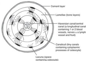
The osteon is the functional unit of compact bone, consisting of concentric layers (lamellae) around a central canal.
Cement Layer: Outermost layer of the osteon.
Lamellae: Concentric bone layers.
Haversian Canal: Central canal containing blood vessels, nerves, and lymph.
Canaliculi: Tiny canals connecting osteocytes.
Lacunae: Spaces containing osteocytes.
Spongy Bone: Lacks osteons; contains trabeculae instead.
Unique Features of Blood and Cartilage
Blood: Unique for its liquid ground substance and soluble fibers in plasma.
Cartilage: Unique for its lack of blood supply and gel-like matrix.
Muscle Tissue Types
Muscle tissue is specialized for movement and classified as:
Skeletal Muscle: Striated, voluntary, cylindrical, multinucleated, attached to bones.
Cardiac Muscle: Striated, involuntary, branched, uninucleated, contains intercalated discs (with desmosomes and gap junctions), found in the heart.
Smooth Muscle: Non-striated, involuntary, spindle-shaped, uninucleated, found in walls of hollow organs and blood vessels.
Nervous Tissue
Neurons: Conduct action potentials; generally do not undergo mitosis.
Neuroglial Cells: Support cells; do not conduct action potentials but can undergo mitosis.
Extracellular Space: Fluid-filled matrix between neurons and neuroglia.
Membranes
Body membranes consist of epithelial and connective tissue components:
Mucous Membranes: Epithelial layer secretes mucus; underlying lamina propria (loose connective tissue); lines respiratory, digestive, and reproductive tracts.
Serous Membranes: Single layer of squamous epithelial cells (mesothelium) with underlying connective tissue; lines body cavities (pericardium, peritoneum, pleura).
Synovial Membranes: Inner synoviocytes produce synovial fluid; outer fibrous layer protects; found in joints.
Cutaneous Membrane (Skin): Epidermis (keratinized stratified squamous epithelium); dermis (areolar and dense irregular connective tissue).
Tissue Repair
Tissue repair involves three main steps:
Inflammation: Trauma triggers release of inflammatory chemicals (histamine, serotonin, prostaglandins); blood vessels become leaky, allowing WBCs and plasma proteins to enter tissue; clotting occurs.
Granulation Tissue Formation: New connective tissue and capillaries grow into the injured area, filling the wound base.
Maturation/Regeneration/Fibrosis: Tissue matures; fibrosis (scarring) may occur if fibers are not restored to original pattern, leading to loss of flexibility and function (e.g., cardiac fibrosis).
Parenchyma: Functional tissue of an organ. Stroma: Supportive tissue (connective tissue, blood vessels, nerves, ducts).
The Integumentary System (Skin)
Functions of Skin
Thermoregulation
Protection
Vitamin D Production: Essential for calcium and phosphorus absorption/retention.
Excretion
Blood Reservoir: Dermis holds 8-10% of total blood at rest.
Structure of the Epidermis
Strata (Layers): Corneum (dead keratinocytes), Granulosum, Spinosum (keratin production begins), Basale (deepest).
Keratinocytes: Produce keratin for waterproofing and abrasion resistance.
Tactile (Merkel) Cells: Touch receptors.
Langerhans Cells: Immune cells (macrophages).
Dermis and Hypodermis
Papillary Dermis: Areolar tissue; contains dermal papillae (ridges that lock epidermis and dermis).
Reticular Dermis: Dense irregular connective tissue.
Hypodermis: Adipose tissue beneath skin; not part of skin; provides insulation, shock absorption, protection.
Skin Pigments
Hemoglobin: In dermal capillaries; gives pink tint.
Carotene: Yellow pigment in stratum corneum and hypodermis.
Melanin: Produced by melanocytes in stratum basale; gives brown tint.
Disorders: Vitiligo (loss of melanocytes), Cyanosis (bluish color due to low oxygen).
Hair and Associated Structures
Hair Shaft: Above skin surface.
Hair Root and Matrix: Below skin; matrix is site of mitosis.
Hair Papillae: Connective tissue nourishing hair root.
Arrector Pili: Smooth muscle causing hair to stand (goosebumps).
Hair Color: Determined by melanin amount.
Glands of the Skin
Sebaceous Glands: Produce sebum (oil) for lubrication; holocrine secretion; found everywhere except palms and soles.
Sudoriferous (Sweat) Glands:
Eccrine: Most numerous, thermoregulation, active throughout life (merocrine secretion).
Apocrine: Activate at puberty, found in axillary/genital regions, responsible for body odor.
Ceruminous Glands: Modified apocrine glands producing earwax (cerumen).
Nails
Composition: Made of keratin.
Growth: Occurs at nail root (matrix).
Color: Pink due to blood supply; pale nails may indicate anemia or disease.
