 Back
BackTissues: Structure, Function, and Clinical Relevance
Study Guide - Smart Notes
 Tailored notes based on your materials, expanded with key definitions, examples, and context.
Chapter 4: Tissues
Introduction to Tissues
Tissues are groups of cells with a common structure and function. The study of tissues, known as histology, is fundamental to understanding how the human body is organized and how it functions in health and disease. There are four primary tissue types: epithelial, connective, muscle, and nervous tissue.
Epithelial Tissue
Special Characteristics of Epithelium
Epithelial tissue covers body surfaces, lines internal cavities, and forms glands. It is characterized by closely packed cells with minimal extracellular material, specialized cell junctions, polarity (distinct apical and basal surfaces), and a basement membrane that anchors it to underlying connective tissue.
Polarity: Apical (top) surface faces the body surface or lumen; basal (bottom) surface attaches to the basement membrane.
Cellularity: Composed almost entirely of tightly packed cells.
Specialized Contacts: Includes tight junctions, desmosomes, and gap junctions for adhesion and communication.
Avascular but Innervated: Contains no blood vessels but is supplied by nerve fibers.
Regeneration: High capacity for renewal due to frequent cell loss and replacement.
Classification of Epithelia
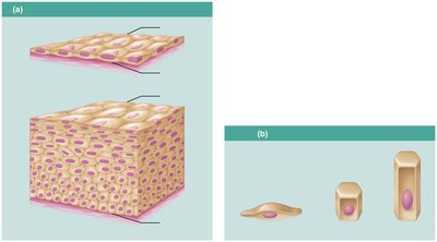
Epithelia are classified by the number of cell layers and the shape of the cells at the apical surface.
Simple epithelium: Single cell layer (for absorption, secretion, filtration).
Stratified epithelium: Multiple layers (for protection).
Cell shapes: Squamous (flat), cuboidal (cube-like), columnar (tall).
Types of Epithelial Tissues
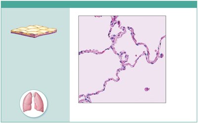
Simple Squamous Epithelium: Single layer of flat cells; allows diffusion and filtration; found in air sacs of lungs, lining of heart, blood vessels, and serosae.
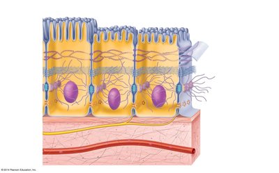
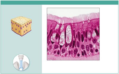
Pseudostratified Columnar Epithelium: Single layer of cells of varying heights; often ciliated; secretes and propels mucus; lines trachea and upper respiratory tract.
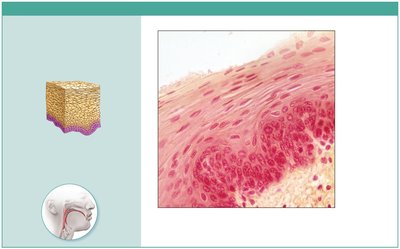
Stratified Squamous Epithelium: Multiple layers; protects against abrasion; nonkeratinized type lines esophagus, mouth, vagina; keratinized type forms epidermis of skin.
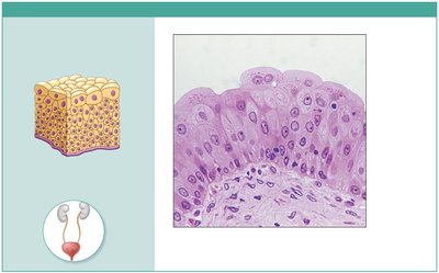
Transitional Epithelium: Resembles both stratified squamous and cuboidal; stretches to permit distension; lines ureters, bladder, and part of urethra.
Cell Junctions in Epithelia
Cell junctions are specialized connections between epithelial cells that maintain tissue integrity and function.
Tight Junctions: Form impermeable seals to prevent passage of molecules between cells.
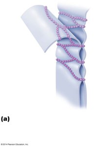
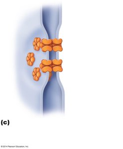
Desmosomes: Anchoring junctions that bind cells together and resist mechanical stress.
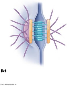
Gap Junctions: Communicating junctions that allow ions and small molecules to pass between cells.
Surface Specializations
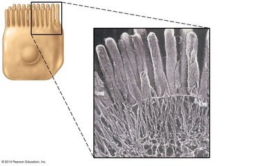
Microvilli: Fingerlike extensions of the plasma membrane that increase surface area for absorption (e.g., in the small intestine).
Cilia: Motile projections that move substances across the epithelial surface (e.g., in the respiratory tract).
Connective Tissue
Overview and Classification
Connective tissue supports, binds, and protects other tissues and organs. It is characterized by cells embedded in an extracellular matrix composed of fibers and ground substance. Connective tissues are classified into connective tissue proper, cartilage, bone, and blood.
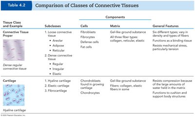
Tissue Class | Subclasses | Cells | Matrix | General Features |
|---|---|---|---|---|
Connective Tissue Proper | Loose (areolar, adipose, reticular); Dense (regular, irregular, elastic) | Fibroblasts, fibrocytes, defense cells, fat cells | Gel-like ground substance; collagen, reticular, elastic fibers | Binding, support, resistance to tension |
Cartilage | Hyaline, elastic, fibrocartilage | Chondroblasts, chondrocytes | Gel-like ground substance; collagen, elastic fibers | Resists compression, supports body structures |
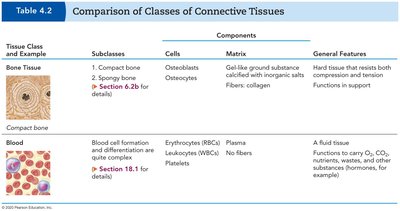
Bone | Compact, spongy | Osteoblasts, osteocytes | Hard, calcified; collagen fibers | Support, protection, storage |
Blood | --- | Erythrocytes, leukocytes, platelets | Plasma (fluid) | Transport of gases, nutrients, wastes |
Areolar Connective Tissue: A Model Connective Tissue
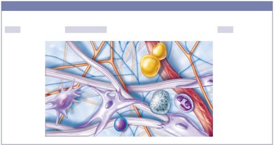
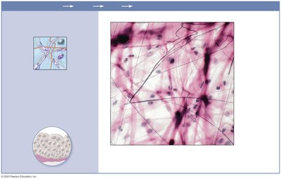
Areolar tissue is a loose connective tissue that serves as a universal packing material between other tissues. It contains a variety of cell types (fibroblasts, macrophages, mast cells, white blood cells) and all three fiber types (collagen, elastic, reticular) in a gel-like ground substance.
Types of Connective Tissue Proper
Loose Areolar: Cushions organs, holds tissue fluid, involved in inflammation.
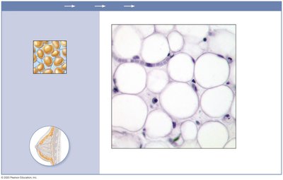
Loose Adipose: Stores fat, insulates, supports and protects organs.
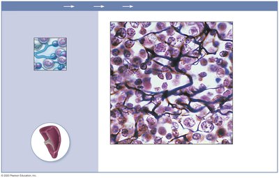
Loose Reticular: Forms a soft internal skeleton (stroma) for lymphoid organs.
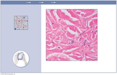
Dense Irregular: Withstands tension in many directions; found in dermis, joint capsules.
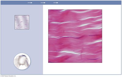
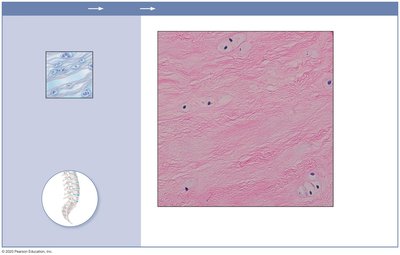
Dense Regular: Parallel collagen fibers; found in tendons and ligaments.
Dense Elastic: High proportion of elastic fibers; found in walls of large arteries.

Cartilage
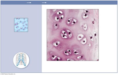
Hyaline Cartilage: Most common; supports, reinforces, cushions; found in embryonic skeleton, ends of long bones, ribs, nose, trachea, larynx.
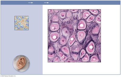
Elastic Cartilage: Maintains shape with flexibility; found in external ear, epiglottis.
Fibrocartilage: Absorbs compressive shock; found in intervertebral discs, pubic symphysis, knee discs.
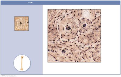
Bone (Osseous Tissue)
Bone tissue is a hard, calcified connective tissue with abundant collagen fibers. It supports and protects body structures, provides levers for movement, stores minerals, and houses marrow for blood cell formation.
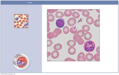
Blood
Blood is a fluid connective tissue composed of red blood cells (erythrocytes), white blood cells (leukocytes), platelets, and plasma. It functions in the transport of gases, nutrients, wastes, and other substances throughout the body.
Summary Table: Epithelial and Connective Tissue Types
Tissue Type | Key Features | Main Locations | Main Functions |
|---|---|---|---|
Simple Squamous Epithelium | Single flat layer | Alveoli, blood vessels | Diffusion, filtration |
Pseudostratified Columnar Epithelium | Single layer, varied height, cilia | Trachea, upper respiratory tract | Mucus secretion, propulsion |
Stratified Squamous Epithelium | Multiple layers, flat surface cells | Skin, mouth, esophagus | Protection |
Transitional Epithelium | Multiple layers, dome-shaped surface cells | Bladder, ureters | Stretching |
Areolar Connective Tissue | Loose, all fiber types | Under epithelia | Cushioning, immunity |
Adipose Tissue | Fat storage | Under skin, around organs | Energy storage, insulation |
Reticular Connective Tissue | Network of reticular fibers | Lymphoid organs | Support |
Dense Regular Connective Tissue | Parallel collagen fibers | Tendons, ligaments | Attachment, tensile strength |
Dense Irregular Connective Tissue | Irregular collagen fibers | Dermis, joint capsules | Strength in many directions |
Elastic Connective Tissue | Elastic fibers | Arteries, bronchial tubes | Recoil |
Hyaline Cartilage | Firm, glassy matrix | Joints, ribs, nose | Support, cushion |
Elastic Cartilage | Elastic fibers | Ear, epiglottis | Flexibility |
Fibrocartilage | Thick collagen fibers | Intervertebral discs | Shock absorption |
Bone | Calcified matrix | Skeletal system | Support, protection |
Blood | Fluid matrix | Blood vessels | Transport |
Clinical Relevance and Application
Understanding tissue structure and function is essential for clinical practice. For example, damage to the keratinized stratified squamous epithelium of the skin (as in burns) compromises the barrier against infection and water loss. Cartilage degeneration in joints leads to pain and impaired mobility, posing challenges for repair due to cartilage's avascular nature.
Key Terms
Epithelium: Tissue forming outer layer of body surfaces and lining cavities.
Basement membrane: Thin layer anchoring epithelium to connective tissue.
Extracellular matrix: Non-cellular material in connective tissue, consisting of fibers and ground substance.
Chondrocyte: Cartilage cell.
Osteocyte: Bone cell.
Erythrocyte: Red blood cell.
Leukocyte: White blood cell.
Example: In a clinical scenario, a second-degree burn damages the epidermis (keratinized stratified squamous epithelium) and part of the dermis, increasing risk of infection and dehydration. Management includes wound care, infection prevention, and monitoring for fluid loss.
Additional info: For further study, review the structure and function of muscle and nervous tissues, as well as the integration of tissues in organs and organ systems.
