 Back
BackTissues: The Living Fabric – Study Notes for Anatomy & Physiology
Study Guide - Smart Notes
 Tailored notes based on your materials, expanded with key definitions, examples, and context.
Tissues: The Living Fabric
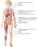
Introduction to Tissues and Histology
Tissues are groups of cells with similar structure and function, forming the foundation of organs and systems in the human body. The study of tissues is known as histology. Understanding tissue types is essential for monitoring health and diagnosing conditions such as bedsores and cancer.
Four Basic Tissue Types: Epithelial, Connective, Muscle, and Nervous tissue
Homeostasis: Each tissue type contributes to maintaining internal balance
Histology: Microscopic study of tissues
Preparation of Tissue Samples for Microscopy
To study tissues under a microscope, samples must be properly prepared. This involves fixing, sectioning, and staining to enhance visibility and contrast.
Fixation: Preserves tissue structure
Sectioning: Thin slices allow light or electrons to pass through
Staining: Dyes or metal salts highlight structures; may introduce artifacts
Microscopy Types: Light microscopy (colored dyes), Electron microscopy (heavy metal salts)
Epithelial Tissue
Definition and Functions
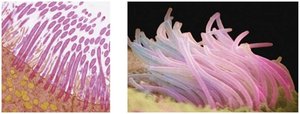
Epithelial tissue (epithelium) forms sheets that cover surfaces or line cavities. It is specialized for protection, absorption, filtration, excretion, secretion, and sensory reception.
Covering and Lining Epithelium: Skin, linings of urogenital, digestive, and respiratory systems
Glandular Epithelium: Forms glands (e.g., salivary glands)
Special Characteristics of Epithelial Tissue
Epithelial tissues possess five key characteristics:
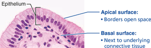
Polarity: Distinct apical (top) and basal (bottom) surfaces
Specialized Contacts: Tight junctions and desmosomes bind cells
Supported by Connective Tissue: Basement membrane reinforces and defines boundaries
Avascular but Innervated: No blood vessels, but supplied by nerves
Regeneration: High capacity for renewal, especially in areas exposed to friction
Classification of Epithelial Tissue
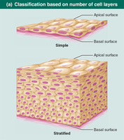
Epithelial tissues are classified by cell layers and cell shape:
Number of Layers:
Simple Epithelium: Single layer, ideal for absorption and filtration
Stratified Epithelium: Multiple layers, suited for protection
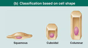
Cell Shape:
Squamous: Flattened, scale-like
Cuboidal: Cube-shaped
Columnar: Tall, column-like
Types of Simple Epithelia
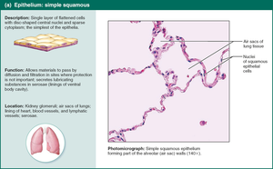
Simple Squamous Epithelium
Single layer of flattened cells, ideal for rapid diffusion and filtration. Found in kidneys, lungs, lining of blood vessels (endothelium), and serous membranes (mesothelium).
Function: Allows materials to pass by diffusion and filtration
Location: Air sacs of lungs, lining of heart, blood vessels
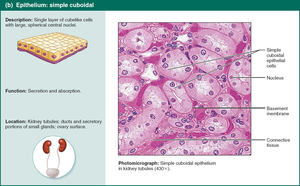
Simple Cuboidal Epithelium
Single layer of cube-shaped cells, specialized for secretion and absorption. Forms walls of kidney tubules and ducts of glands.
Function: Secretion and absorption
Location: Kidney tubules, ducts of small glands
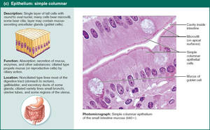
Simple Columnar Epithelium
Single layer of tall cells, often with microvilli or cilia. Contains goblet cells for mucus secretion. Found in digestive tract, gallbladder, bronchi, and uterine tubes.
Function: Absorption, secretion of mucus and enzymes
Location: Digestive tract, uterine tubes
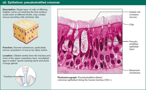
Pseudostratified Columnar Epithelium
Appears multi-layered but is actually a single layer. Often ciliated and contains goblet cells. Found in upper respiratory tract and ducts of large glands.
Function: Secretion and movement of mucus
Location: Trachea, upper respiratory tract
Summary Table: Epithelial Tissue Types
Type | Structure | Function | Location |
|---|---|---|---|
Simple Squamous | Single layer, flat cells | Diffusion, filtration | Lungs, blood vessels |
Simple Cuboidal | Single layer, cube cells | Secretion, absorption | Kidney tubules, glands |
Simple Columnar | Single layer, tall cells | Absorption, secretion | Digestive tract, uterine tubes |
Pseudostratified Columnar | Single layer, varying height | Mucus secretion, movement | Respiratory tract |
Additional info:
Stratified epithelia provide greater protection and are found in areas subject to abrasion (e.g., skin, mouth lining).
Transitional epithelium lines urinary organs and can stretch to accommodate fluctuating volumes.
Glandular epithelia are classified as endocrine (ductless, hormone-secreting) or exocrine (ducts, secrete onto surfaces).
