 Back
BackUnderstanding Cardiac Output and Anatomical Identification
Study Guide - Smart Notes
 Tailored notes based on your materials, expanded with key definitions, examples, and context.
Q1. What is the definition of Cardiac Output (CO)?
Background
Topic: Cardiovascular Physiology
This question tests your understanding of cardiac output, a fundamental concept in human physiology related to the function of the heart and circulatory system.
Key Terms and Formulas
Cardiac Output (CO): The volume of blood pumped by each ventricle in one minute.
Stroke Volume (SV): The amount of blood pumped out of each ventricle during one contraction.
Heart Rate (HR): The number of heart beats per minute.
The formula for cardiac output is:
Step-by-Step Guidance
Review the definitions of each option provided in the question. Pay attention to the terms "ventricle," "minute," and "contraction."
Recall that cardiac output is a measure of blood flow, not just heart rate or electrical impulses.
Understand that cardiac output is calculated by multiplying the stroke volume (amount of blood pumped per beat) by the heart rate (beats per minute).
Eliminate options that refer only to heart rate, electrical impulses, or blood volume at a single point in the cardiac cycle.
Try solving on your own before revealing the answer!
Final Answer: The amount of blood pumped out by each ventricle in 1 minute
Cardiac output is defined as the volume of blood pumped by each ventricle per minute. It is a key indicator of heart function and is calculated as stroke volume multiplied by heart rate.
Q2. Identify the anatomical structures labeled A, B, C, D, and E in the provided cadaver image.
Background
Topic: Human Anatomy – Thoracic and Abdominal Structures
This question tests your ability to recognize and name anatomical structures in a cadaveric specimen, a skill essential for practical anatomy exams.
Key Terms
Thoracic cavity: Contains the heart and lungs.
Diaphragm: Muscle separating thoracic and abdominal cavities.
Major vessels: Such as the aorta and vena cava.
Step-by-Step Guidance
Observe the orientation of the cadaver: note the head, thorax, and abdomen.
Identify the labeled structures based on their location and relation to other visible organs.
Recall the major anatomical landmarks in the thoracic and abdominal regions.
Use your knowledge of anatomy to match each label (A, B, C, D, E) to the correct structure.
Try solving on your own before revealing the answer!
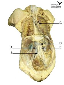
Final Answer:
A: Right lung
B: Heart
C: Left lung
D: Diaphragm
E: Liver
These are the major structures visible in the thoracic and upper abdominal regions of the cadaver.
