 Back
BackUnderstanding the Structure of the Human Heart
Study Guide - Smart Notes
 Tailored notes based on your materials, expanded with key definitions, examples, and context.
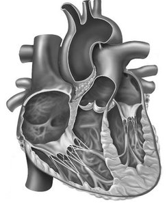
Q: Study the anatomy of the human heart as shown in the image. Identify the major chambers, valves, and vessels visible in this sectioned view.
Background
Topic: Cardiovascular System – Heart Anatomy
This question is testing your ability to recognize and name the main anatomical features of the heart, including its chambers, valves, and associated blood vessels, as seen in a cross-sectional diagram.
Key Terms and Concepts:
Atria: The two upper chambers of the heart (right and left atrium) that receive blood returning to the heart.
Ventricles: The two lower chambers (right and left ventricle) that pump blood out of the heart.
Valves: Structures that prevent backflow of blood (e.g., tricuspid, bicuspid/mitral, pulmonary, and aortic valves).
Major Vessels: Include the aorta, pulmonary arteries, pulmonary veins, and vena cavae.
Step-by-Step Guidance
Begin by identifying the four chambers of the heart in the image: right atrium, right ventricle, left atrium, and left ventricle. Notice the thickness of the walls—left ventricle walls are typically the thickest.
Locate the major valves between the chambers and at the exits of the heart: the tricuspid valve (between right atrium and right ventricle), the bicuspid/mitral valve (between left atrium and left ventricle), the pulmonary valve (leading to the pulmonary artery), and the aortic valve (leading to the aorta).
Identify the large blood vessels entering and leaving the heart: the superior and inferior vena cava (bringing blood to the right atrium), the pulmonary trunk/arteries (carrying blood from the right ventricle to the lungs), the pulmonary veins (bringing blood from the lungs to the left atrium), and the aorta (carrying blood from the left ventricle to the body).
Observe the direction of blood flow through the heart: from atria to ventricles, then out through the arteries.
Try solving on your own before revealing the answer!
Final Answer:
The image shows a sectioned human heart with the right and left atria at the top, right and left ventricles at the bottom, the tricuspid and bicuspid (mitral) valves between the atria and ventricles, and the aortic and pulmonary valves at the exits. The aorta, pulmonary trunk, and vena cavae are also visible as major vessels connected to the heart.
Recognizing these structures is essential for understanding the flow of blood through the heart and the function of each chamber and valve.
