 Back
Unit 1: Introduction to Human Anatomy and Physiology – Structured Study Notes
Study Guide - Smart Notes
 Tailored notes based on your materials, expanded with key definitions, examples, and context.
Introduction to Anatomy & Physiology
Definition and Distinction
Anatomy and physiology are foundational sciences that explain the structure and function of the human body. Anatomy is the study of body structure, while physiology focuses on the mechanisms and processes that allow these structures to function.
Systemic Anatomy: Study of individual organ systems (e.g., cardiovascular system).
Regional Anatomy: Study of specific regions (e.g., head, neck).
Surface Anatomy: Study of external body markings.
Gross Anatomy: Study of structures visible to the naked eye.
Microscopic Anatomy: Study of structures requiring magnification (e.g., histology, cytology).
Physiology: Study of functional processes, including neurophysiology and cardiovascular physiology.
Core Insight: Anatomy provides the blueprint; physiology provides the action.
Characteristics of Life
Seven Key Traits
All living organisms share seven interconnected characteristics that define life:
Cellular Composition: Cells are the basic unit of life.
Metabolism: Chemical processes that build (anabolism) and break down (catabolism) molecules to produce ATP.
Growth: Increase in size when anabolism exceeds catabolism.
Excretion: Removal of metabolic waste products.
Responsiveness: Ability to sense and react to stimuli.
Movement: Motion in response to stimuli (e.g., muscle contraction, blood flow).
Reproduction: Cell division for growth/repair and production of offspring.
Structural Levels of Organization
Six Major Levels
The human body is organized hierarchically from simple to complex:
Chemical: Atoms and molecules (e.g., water, DNA).
Cellular: Molecules form cells (e.g., muscle cell).
Tissue: Groups of similar cells and extracellular matrix (ECM) working together.
Organ: Two or more tissues form a structural unit (e.g., heart).
Organ System: Groups of organs cooperate to meet body needs.
Organism: All organ systems function together to maintain life.
Spatial Terminology
Anatomical Position and Directional Terms
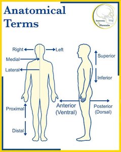
The anatomical position is the standard reference for describing locations and directions in the body: standing upright, feet flat, arms at sides, palms forward.
Right/Left: Always refer to the patient's perspective.
Anterior (Ventral)/Posterior (Dorsal): Front/back of the body.
Superior/Inferior: Toward head/toward feet.
Proximal/Distal: Closer/farther from point of origin.
Medial/Lateral: Closer/farther from midline.
Superficial/Deep: Closer/farther from body surface.
Sectional Planes
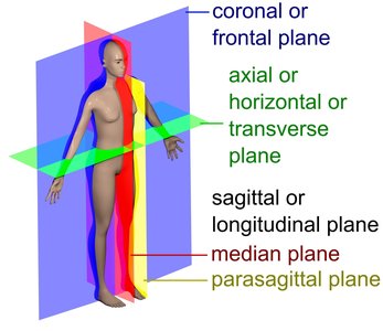
Three primary planes are used to slice the body for anatomical study:
Sagittal Plane: Divides body into right and left portions. Midsagittal is exactly at midline; parasagittal is parallel but off-center.
Frontal (Coronal) Plane: Divides body into anterior and posterior portions.
Transverse (Axial/Horizontal) Plane: Divides body into superior and inferior portions.
Body Cavities
Major Cavities and Their Subdivisions
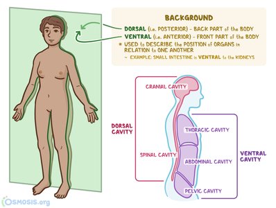
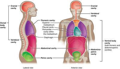
The body contains two major cavities that protect and organize internal organs:
Dorsal Body Cavity: Posterior; includes cranial cavity (brain) and vertebral/spinal cavity (spinal cord).
Ventral Body Cavity: Anterior; separated by diaphragm, includes thoracic cavity (lungs, heart) and abdominopelvic cavity (digestive, reproductive, urinary organs).
Thoracic and Abdominopelvic Cavities
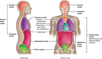
The ventral cavity is further divided:
Thoracic Cavity: Contains pleural cavities (lungs), mediastinum (trachea, esophagus, major vessels), and pericardial cavity (heart).
Abdominopelvic Cavity: Contains abdominal cavity (digestive organs) and pelvic cavity (reproductive organs, bladder).
Diaphragm: Skeletal muscle separating thoracic and abdominopelvic cavities.
Abdominopelvic Quadrants and Regions
Four Quadrants
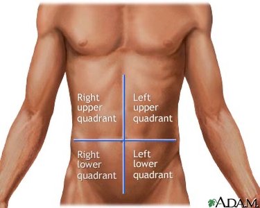
The abdominopelvic area is divided into four clinical quadrants for diagnostic purposes:
Right Upper Quadrant (RUQ): Liver, gallbladder.
Left Upper Quadrant (LUQ): Stomach, spleen.
Right Lower Quadrant (RLQ): Appendix, cecum.
Left Lower Quadrant (LLQ): Colon, sigmoid colon.
Nine Anatomical Regions
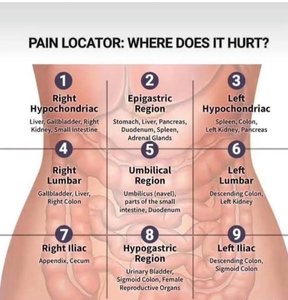
For more precise mapping, the abdominopelvic area is divided into nine regions:
Top Row: Right Hypochondriac, Epigastric, Left Hypochondriac.
Middle Row: Right Lumbar, Umbilical, Left Lumbar.
Bottom Row: Right Iliac (Inguinal), Hypogastric (Pubic), Left Iliac (Inguinal).
Serous Membranes
Structure and Function
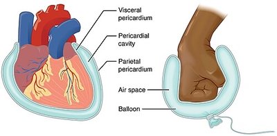
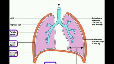
Serous membranes are double-layered structures that reduce friction between organs and cavity walls:
Parietal Layer: Lines cavity walls.
Visceral Layer: Covers organs directly.
Serous Cavity: Fluid-filled space between layers, lubricated by serous fluid.
Three key serous membranes:
Pleura: Lungs.
Pericardium: Heart.
Peritoneum: Abdominal organs.
Homeostasis
Principle and Mechanisms
Homeostasis is the maintenance of a stable internal environment. It involves dynamic balance around set points for variables like temperature, pH, and glucose.
Regulated Variables: Must be kept within narrow ranges.
Imbalances: Lead to illness or death.
Continuous Correction: Metabolic energy is used to monitor and restore balance.
Feedback Loops
Homeostasis is maintained by feedback loops:
Negative Feedback: Counteracts changes, returning variables to set points. Example: Thermoregulation (shivering when cold).
Positive Feedback: Amplifies changes until a process is completed. Example: Blood clotting, childbirth contractions.
Steps in a feedback loop:
Stimulus: Change detected.
Receptor: Sensor detects change.
Control Center: Compares to set point, decides action.
Effector: Carries out response to restore balance.
Principles of Complementarity and Gradients
Form Complements Function
Structure is always designed for function. For example, thin alveolar walls in the lungs allow rapid gas exchange; thickened walls (fibrosis) impede this process.
Gradients and Cell Communication
Movement in the body is driven by gradients (temperature, concentration, pressure). Cells communicate via electrical and chemical signals to coordinate activities and maintain homeostasis.
Active Recall and Clinical Application
Sample Questions
What major skeletal muscle separates the thoracic and abdominopelvic cavities? Diaphragm
Shivering in response to cold is an example of what kind of feedback loop? Negative feedback
Which plane of section slices the body into anterior and posterior portions? Frontal (coronal) plane
What fluid fills the space between parietal and visceral membranes? Serous fluid
Key Equations
ATP Production (Metabolism)
Cellular respiration equation:
Homeostatic Feedback Loop (Generalized)
Feedback loop schematic:
Summary Table: Directional Terms
Term | Definition | Example |
|---|---|---|
Anterior | Toward the front | Heart is anterior to spine |
Posterior | Toward the back | Spinal cord is posterior to esophagus |
Superior | Toward the head | Nose is superior to mouth |
Inferior | Toward the feet | Pelvis is inferior to ribcage |
Proximal | Closer to origin | Elbow is proximal to wrist |
Distal | Farther from origin | Toes are distal to ankle |
Medial | Closer to midline | Sternum is medial to shoulder |
Lateral | Farther from midline | Ears are lateral to nose |
Superficial | Closer to surface | Skin is superficial to muscle |
Deep | Farther from surface | Bone is deep to skin |
Additional info: Academic context and expanded explanations were added to ensure completeness and clarity for exam preparation.
