 Back
BackUnit 1 Practical Study Guide: Anatomical Terminology, Epithelial and Connective Tissues
Study Guide - Smart Notes
 Tailored notes based on your materials, expanded with key definitions, examples, and context.
Anatomical Terminology and Landmarks
Overview of Anatomical Terminology
Anatomical terminology provides a standardized language for describing the locations and relationships of body parts. This system is used worldwide to ensure clear communication among healthcare professionals and scientists.
Noun forms refer to the structure itself (e.g., acromion for the shoulder).
Adjective forms describe the region or related structures (e.g., acromial for the shoulder region).
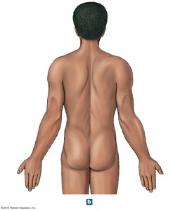
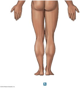
Posterior Anatomical Landmarks
The posterior (dorsal) view of the body highlights several key anatomical regions:
Acromial: Shoulder
Dorsal: Back
Olecranal: Back of elbow
Lumbar: Loin (lower back)
Cervical: Neck
Cephalic: Head
Gluteal: Buttock
Popliteal: Back of knee
Sural: Calf
Calcaneal: Heel of foot
Plantar: Sole of foot
Epithelial Tissue
Specializations of Epithelial Cells
Epithelial cells may possess surface specializations that enhance their function:
Microvilli: Increase surface area for absorption, commonly found in the intestines and kidney tubules.
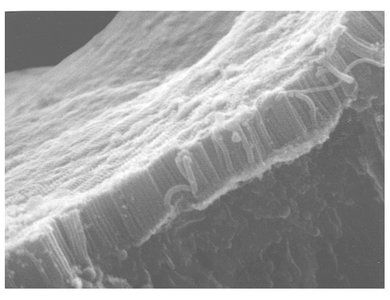
Cilia: Motile projections that move substances across the epithelial surface, such as in the respiratory tract.
Types of Epithelial Tissue
Epithelial tissues are classified based on the number of cell layers and the shape of the cells at the apical surface.
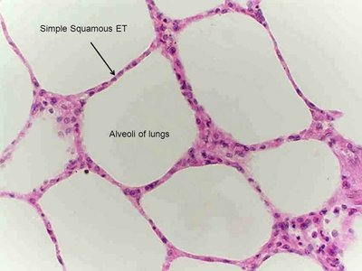
Simple Squamous Epithelium
Structure: Single layer of flat cells.
Function: Facilitates diffusion and filtration.
Locations: Alveoli of lungs, lining of blood vessels, serous membranes.
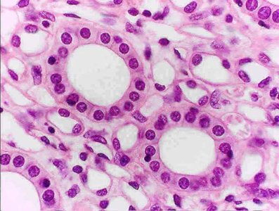
Simple Cuboidal Epithelium
Structure: Single layer of cube-shaped cells.
Function: Secretion and absorption.
Locations: Kidney tubules, glandular ducts, thyroid gland.
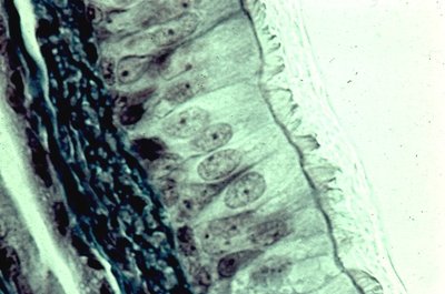
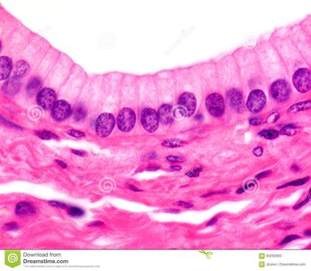
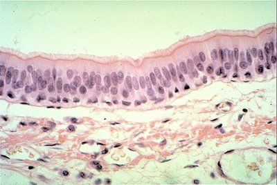
Simple Columnar Epithelium
Structure: Single layer of tall, column-like cells.
Function: Absorption and secretion; may have microvilli or cilia.
Locations: Lining of stomach, small intestine, and large intestine.
Stratified Cuboidal Epithelium
Structure: Multiple layers of cube-shaped cells.
Function: Protection, secretion, and absorption.
Locations: Ducts of sweat glands, mammary glands, and salivary glands.
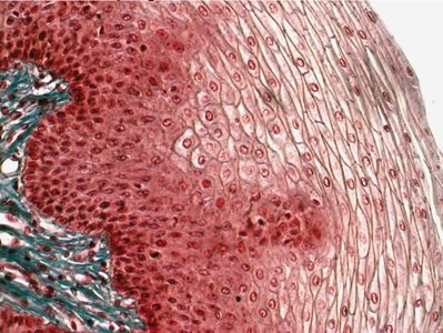
Stratified Squamous Epithelium
Structure: Multiple layers with flat cells at the surface.
Function: Protects underlying tissues from abrasion.
Locations: Epidermis of skin, lining of mouth, esophagus, and vagina.
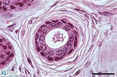
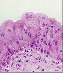
Pseudostratified Ciliated Columnar Epithelium
Structure: Appears stratified but all cells contact the basement membrane; often ciliated.
Function: Secretion and movement of mucus.
Locations: Lining of the trachea and upper respiratory tract.
Transitional Epithelium
Structure: Multiple layers of cells that can change shape (from cuboidal to squamous).
Function: Allows for stretching and distension.
Locations: Urinary bladder, ureters, and part of the urethra.
Connective Tissue Proper
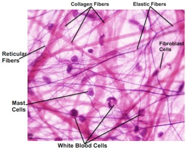
Areolar Connective Tissue
Areolar tissue is a loose connective tissue that provides support and flexibility to organs and tissues.
Components: Collagen fibers, elastic fibers, reticular fibers, fibroblasts, mast cells, and white blood cells.
Function: Cushions organs, provides support but permits independent movement, and defends against pathogens.
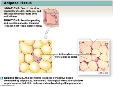
Adipose Tissue
Adipose tissue is a specialized loose connective tissue that stores fat.
Location: Deep to the skin, around organs, in the buttocks, breasts, and padding around eyes and kidneys.
Function: Provides padding, cushions shocks, insulates (reduces heat loss), and stores energy.
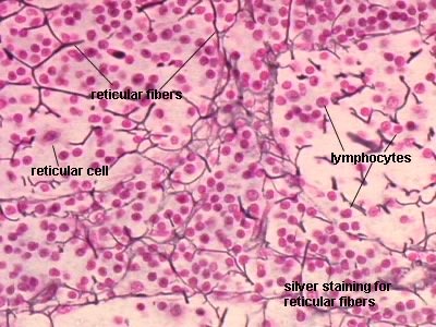
Reticular Tissue
Reticular tissue forms a supporting framework for soft organs such as the liver, bone marrow, and lymphatic organs.
Components: Reticular fibers and reticular cells.
Function: Provides a supportive mesh for cells in organs.
Dense Regular Connective Tissue
Dense regular connective tissue is composed of parallel collagen fibers and provides strong attachment between structures.
Location: Tendons, most ligaments, aponeuroses.
Function: Withstands great tensile stress when pulling force is applied in one direction.
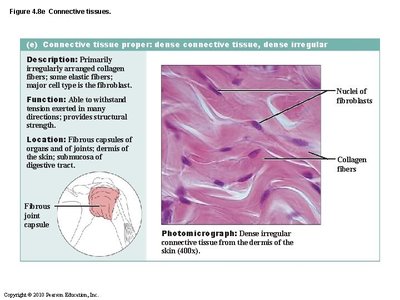
Dense Irregular Connective Tissue
Dense irregular connective tissue contains collagen fibers arranged in multiple directions, providing strength in many directions.
Location: Dermis of the skin, fibrous capsules of organs and joints, submucosa of digestive tract.
Function: Withstands tension exerted in many directions; provides structural strength.
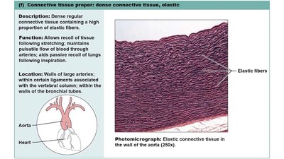
Dense Elastic Connective Tissue
Dense elastic connective tissue contains a high proportion of elastic fibers, allowing tissues to recoil after stretching.
Location: Walls of large arteries, certain ligaments in the vertebral column, walls of bronchial tubes.
Function: Allows recoil of tissue following stretching; maintains pulsatile flow of blood through arteries.
Summary Table: Epithelial Tissue Types
Type | Structure | Function | Location |
|---|---|---|---|
Simple Squamous | Single layer, flat cells | Diffusion, filtration | Alveoli, blood vessels |
Simple Cuboidal | Single layer, cube-shaped | Secretion, absorption | Kidney tubules, glands |
Simple Columnar | Single layer, tall cells | Absorption, secretion | Digestive tract |
Stratified Squamous | Multiple layers, flat surface cells | Protection | Skin, mouth, esophagus |
Pseudostratified Ciliated Columnar | Single layer, appears stratified, cilia | Secretion, movement of mucus | Respiratory tract |
Transitional | Multiple layers, variable shape | Stretching | Urinary bladder |
Additional info: The above content covers foundational anatomical terminology, epithelial tissue types, and connective tissue proper, which are essential for understanding human anatomy and physiology at the college level.
