 Back
BackUnit 6: The Respiratory System – Structure, Function, and Regulation
Study Guide - Smart Notes
 Tailored notes based on your materials, expanded with key definitions, examples, and context.
Functions of the Respiratory System
Overview of Respiratory Functions
The respiratory system is essential for gas exchange, speech, olfaction, regulation of blood pH, and assisting the flow of lymph and venous blood. Its primary function is to supply oxygen (O2) to the body and remove carbon dioxide (CO2).
Gas Exchange: O2 is absorbed, and CO2 is expelled.
Speech (Vocalization): Air movement across vocal cords produces sound.
Smell: Olfactory receptors in the nasal cavity detect odors.
Blood pH Regulation: By controlling CO2 levels, the respiratory system helps maintain acid-base balance.
Flow of Lymph and Venous Blood: Respiratory movements assist in the return of blood and lymph to the heart.

Anatomy of the Respiratory System
Major Structures and Their Functions
The respiratory tract is divided into upper and lower regions, each with specialized structures for air conduction, filtration, and gas exchange.
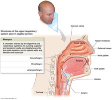
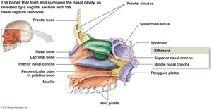
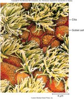
Nasal Conchae: Mucosa-covered projections that increase surface area and turbulence, warming and moistening incoming air. Cilia on the mucosa trap particulate matter.
Palate: Divided into hard (bony) and soft (muscular) palate, separating the nasal and oral cavities.
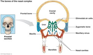
Sinuses: Air-filled spaces (frontal, sphenoidal, maxillary, ethmoidal) that lighten the skull and help warm and moisten air.
Uvula: Hangs from the soft palate; closes off the nasal cavity during swallowing to prevent food from entering the nasal passages.
Pharynx: The throat; connects the nasal cavity and mouth to the larynx and esophagus.
Larynx: The voice box; contains vocal cords and is involved in sound production and airway protection.
Glottis: The opening between the vocal cords, allowing air passage into the larynx.
Trachea: The windpipe; lined with cilia and mucus to trap and move foreign particles away from the lungs.
Carina: The last tracheal cartilage where the trachea divides into the primary bronchi.
Bronchi and Bronchioles: The bronchi branch into smaller bronchioles, conducting air deeper into the lungs.
Alveoli: Tiny air sacs surrounded by capillaries; the primary site of gas exchange. Each alveolus is one cell layer thick to facilitate diffusion.
Lungs: The right lung has three lobes (superior, middle, inferior), and the left lung has two lobes (superior, inferior).
Protective Mechanisms of the Respiratory System
Defense Mechanisms
The respiratory system employs several protective mechanisms to prevent the entry of pathogens and debris.
Nose: Nasal hairs, mucus, and cilia trap and remove particles.
Ciliary Escalator: Cilia move mucus and trapped particles toward the pharynx for removal.
Larynx: The epiglottis and vocal folds prevent food and liquids from entering the airway.
Bronchoconstriction: Smooth muscle constriction of bronchioles limits the entry of harmful substances.
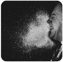
Coughing and Sneezing: Forceful expulsion of air clears irritants from the respiratory tract (irritation reflex).
Macrophages and Lymphocytes: Immune cells in the alveoli and lymph nodes remove debris and pathogens that reach the lower respiratory tract.
Phonation and Voice Production
Mechanisms of Sound Production
Voice production involves the vibration of the vocal cords as air passes over them. The larynx, vocal cords, and articulatory structures (lips, tongue) modify the sound to produce speech.
Laryngeal Cartilages: Include the thyroid, cricoid, epiglottis, and arytenoid cartilages, which support and move the vocal cords.
True Vocal Cords: Vibrate to produce sound; the vestibular (false) vocal folds do not produce sound but help close the glottis.
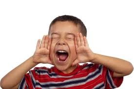
Volume: Increased airflow increases the volume of sound.
Brain Centers: The precentral gyrus initiates voluntary movement; Broca’s area controls motor speech.

Mechanics of Ventilation
Inspiration and Expiration
Ventilation is the process of moving air in and out of the lungs, driven by changes in thoracic volume and pressure.
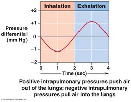
Pressure-Volume Relationship: According to Boyle’s Law, an increase in thoracic volume decreases intrapulmonary pressure, causing air to flow in. Conversely, a decrease in volume increases pressure, causing air to flow out.
Muscles of Inspiration: Diaphragm and external intercostals contract to increase thoracic volume.
Muscles of Expiration: Normal expiration is passive; forced expiration uses internal intercostals and abdominal muscles.
Innervation: The phrenic nerve stimulates the diaphragm; intercostal nerves stimulate the intercostal muscles.
Intrapleural Pressure: The pressure between the lungs and chest wall, always slightly negative to keep lungs inflated.


Control of Breathing
Nervous System Regulation
Rhythmic breathing is controlled by the brainstem, with input from chemoreceptors and stretch receptors.
Pons: Acts as a pacemaker for breathing rhythm.
Medulla Oblongata: Contains respiratory centers that regulate the rate and depth of breathing.
Stretch Receptors: Inhibit inspiration to prevent over-inflation of the lungs.
Phrenic and Intercostal Nerves: Stimulate the diaphragm and intercostal muscles.
Gas Exchange and Transport
Oxygen and Carbon Dioxide Movement
Gas exchange occurs by diffusion across the alveolar-capillary membrane, driven by partial pressure gradients.
Oxygen Transport: 98% bound to hemoglobin (Hb) as oxyhemoglobin; 2% dissolved in plasma.
Carbon Dioxide Transport: 70% as bicarbonate ion in plasma, 23% bound to Hb (carbaminohemoglobin), 7% dissolved in plasma.
Partial Pressure (Dalton’s Law): The total pressure of a gas mixture is the sum of the partial pressures of each component gas.
Key Equation:
This equation explains how CO2 levels affect blood pH.
Respiratory Volumes and Capacities
Definitions and Examples
Tidal Volume (TV): Air moved in and out with each breath at rest.
Inspiratory Reserve Volume (IRV): Additional air inhaled after a normal inspiration.
Expiratory Reserve Volume (ERV): Additional air exhaled after a normal expiration.
Residual Volume (RV): Air remaining in lungs after maximal exhalation.
Vital Capacity (VC): TV + IRV + ERV; maximum air exhaled after maximum inhalation.
Total Lung Capacity (TLC): TV + IRV + ERV + RV; total volume of the lungs.
Control of Ventilation by Blood Gases
Chemoreceptor Regulation
Chemoreceptors monitor blood levels of CO2, O2, and pH to adjust ventilation rate.
Central Chemoreceptors: Located in the medulla; respond to CO2/H+ levels.
Peripheral Chemoreceptors: Located in aortic and carotid bodies; respond to O2 and pH changes.
Hyperventilation: Triggered by increased CO2 or decreased O2/pH.
Hypoventilation: Triggered by decreased CO2 or increased pH.
Pulmonary Diseases
Common Respiratory Disorders
Pulmonary Edema: Fluid accumulation in alveoli, often due to left heart failure.
Emphysema: Destruction of alveolar walls, reducing surface area for gas exchange.
Asthma: Bronchiole constriction and increased mucus production, leading to airflow obstruction.
Pneumothorax: Air in the intrapleural space, causing lung collapse.
Atelectasis: Collapse of part or all of a lung.
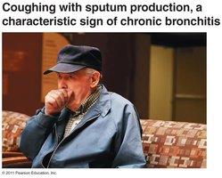
Bronchitis: Inflammation and excess mucus in the bronchi, often with productive cough.
Pneumonia: Infection (often bacterial) causing alveoli to fill with fluid.
Key Terms and Concepts
Definitions
Surfactant: Fluid lining alveoli that reduces surface tension, preventing alveolar collapse.
Ventilation vs. Respiration: Ventilation is air movement; respiration is gas exchange and utilization.
Hypoxia: Inadequate O2 delivery to tissues.
Ischemic Hypoxia: Reduced O2 due to decreased blood flow.
Histotoxic Hypoxia: Tissues cannot use O2 due to cellular damage.
Anemic Hypoxia: Decreased O2 carrying capacity due to low hemoglobin.
Hypoxic Hypoxia: Low arterial PO2 (e.g., high altitude).
