 Back
BackUrinary and Reproductive Systems: Lab Study Guide
Study Guide - Smart Notes
 Tailored notes based on your materials, expanded with key definitions, examples, and context.
Urinary System
Overview of the Urinary System
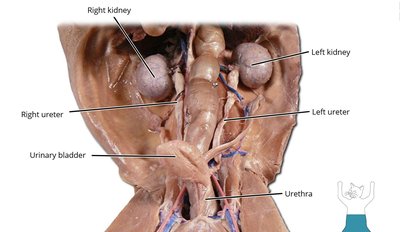
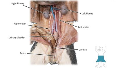
The urinary system is responsible for filtering blood, removing waste products, and regulating fluid and electrolyte balance. It consists of the kidneys, ureters, urinary bladder, and urethra.
Kidneys: Bean-shaped organs that filter blood and produce urine.
Ureters: Muscular tubes that transport urine from the kidneys to the urinary bladder.
Urinary Bladder: A muscular sac that stores urine until it is excreted.
Urethra: The tube through which urine exits the body.
Nephron
The nephron is the functional unit of the kidney, responsible for filtering blood and forming urine.
Glomerulus: A network of capillaries where filtration occurs.
Renal Tubule: Includes the proximal convoluted tubule, loop of Henle, distal convoluted tubule, and collecting duct.
Function: Filtration, reabsorption, secretion, and excretion.
Additional info: Each kidney contains about 1 million nephrons.
Urinalysis
Urinalysis is a diagnostic test that examines the physical, chemical, and microscopic properties of urine.
Physical properties: Color, clarity, and odor.
Chemical properties: pH, protein, glucose, ketones, and urochrome.
Microscopic properties: Cells, crystals, and microorganisms.
Urochrome: The pigment responsible for the yellow color of urine.
Normal pH of Urine: Typically ranges from 4.5 to 8.0, with an average around 6.0.
Urothelium
The urothelium is the transitional epithelium lining the urinary tract, including the renal pelvis, ureters, bladder, and part of the urethra. It allows for stretching and protection against urine toxicity.
Renal Fascia
The renal fascia is a layer of connective tissue that anchors the kidneys to surrounding structures and helps maintain their position in the abdominal cavity.
Histology of the Kidney and Urinary Bladder
Microscopic examination reveals:
Kidney: Renal cortex (contains glomeruli), renal medulla (contains tubules and collecting ducts).
Urinary Bladder: Lined by transitional epithelium (urothelium), with a thick muscular wall (detrusor muscle).
Identification of Cat Urinary System Organs
In laboratory dissections, students should be able to identify the kidneys, ureters, urinary bladder, and urethra in the cat model.

Male Reproductive System
Overview of the Male Reproductive System
The male reproductive system produces, stores, and delivers sperm. Major organs include the testes, epididymis, vas deferens, seminal vesicles, prostate gland, penis, and scrotum.
Testes (Gonads): Produce sperm and testosterone.
Epididymis: Stores and matures sperm.
Vas Deferens: Transports sperm from the epididymis to the urethra.
Spermatic Cord: Contains vas deferens, blood vessels, nerves, and lymphatics.
Penis: Organ of copulation and urination.
Scrotum: Sac that holds and protects the testes.
Prostate Gland and Bulbourethral Glands: Contribute fluids to semen.

Circumcision
Circumcision is the surgical removal of the foreskin (prepuce) from the penis. It is performed for cultural, religious, or medical reasons.
Copulation
Copulation refers to sexual intercourse, during which sperm is deposited into the female reproductive tract.
Identification of Male Cat Reproductive Organs
Students should be able to identify the testes, epididymis, vas deferens, spermatic cord, penis, and scrotum in the male cat dissection.
Identification of Human Male Internal Reproductive Organs
Key internal organs include the testes, epididymis, vas deferens, seminal vesicles, prostate gland, and urethra. (Refer to figure 26.3 in your textbook for detailed anatomy.)
Female Reproductive System
Overview of the Female Reproductive System
The female reproductive system produces ova (eggs), supports fertilization, and houses the developing fetus. Major organs include the ovaries, uterine tubes, uterus, vagina, and external genitalia.
Ovaries (Gonads): Produce ova and hormones (estrogen, progesterone).
Uterine Tubes (Fallopian Tubes): Transport ova from the ovaries to the uterus; site of fertilization.
Uterus: Muscular organ where implantation and fetal development occur. Parts include the fundus, body, and cervix.
Vagina: Muscular canal for copulation and childbirth.
Urogenital Sinus: In some mammals (e.g., cats), a common passage for urinary and reproductive tracts.

Identification of Female Cat Reproductive Organs
Students should be able to identify the ovaries, uterine tubes, uterine horns, uterine body, vagina, urethra, and urogenital sinus in the female cat dissection.

Identification of Human Female Internal Reproductive Organs
Key internal organs include the ovaries, uterine tubes, uterus, and vagina. (Refer to figures 26.10a and 26.11 in your textbook for detailed anatomy.)
Laboratory Procedures and Activities
Urinalysis Lab Procedure
Students should review the steps for performing urinalysis, including collection, observation, chemical testing (dipsticks), and microscopic examination.
STD Epidemic Procedure
This activity models the spread of sexually transmitted diseases (STDs) in a population, emphasizing the importance of prevention and safe practices.
Summary Table: Major Organs of the Urinary and Reproductive Systems
Organ | System | Main Function |
|---|---|---|
Kidney | Urinary | Filtration of blood, urine formation |
Ureter | Urinary | Transport urine to bladder |
Urinary Bladder | Urinary | Stores urine |
Urethra | Urinary/Reproductive (male) | Excretes urine; conducts semen (male) |
Testes | Reproductive (male) | Produce sperm and testosterone |
Ovaries | Reproductive (female) | Produce ova and hormones |
Uterus | Reproductive (female) | Supports fetal development |
Penis | Reproductive (male) | Copulation, semen delivery |
Vagina | Reproductive (female) | Copulation, birth canal |
