 Back
BackUrinary System: Anatomy and Physiology Study Guide
Study Guide - Smart Notes
 Tailored notes based on your materials, expanded with key definitions, examples, and context.
Urinary System Overview
Organs of the Urinary System
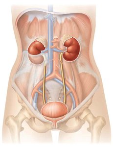
The urinary system is responsible for filtering blood, removing waste, and regulating fluid and electrolyte balance. It consists of four main organs:
Kidneys: Filter blood and produce urine.
Ureters: Transport urine from kidneys to bladder.
Urinary Bladder: Stores urine until excretion.
Urethra: Conducts urine from bladder to exterior.
Functions of the Kidneys
Filtration: Kidneys filter 180-200L of blood daily.
Hormone Production: Produce hormones such as erythropoietin and renin.
Regulation of Blood Chemistry: Control ionic composition, pH, volume, pressure, and osmolarity.
Blood Glucose Regulation: Assist in maintaining blood glucose levels.
Excretion of Metabolic Wastes: Remove urea, creatinine, and other wastes.
Anatomy of the Kidneys
External Structure and Location
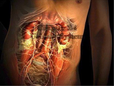
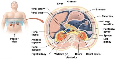
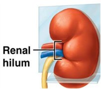
The kidneys are retroperitoneal organs located in the abdominopelvic cavity, partially protected by the 11th and 12th ribs. The medial border features the renal hilum, which serves as the entry and exit point for ureters, blood vessels, lymphatics, and nerves.
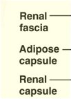
Support Structures and Coverings
Renal fascia: Anchors the kidney to surrounding tissues.
Adipose capsule: Cushions and protects the kidney.
Renal capsule: Thin, tough layer directly covering the kidney.
Internal Anatomy of the Kidney
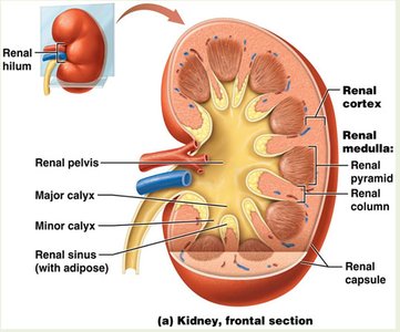
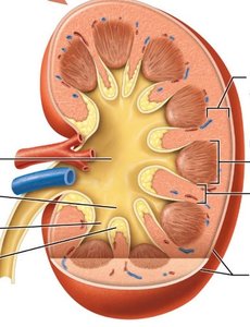
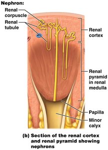
The functional portion of the kidney includes the renal cortex and renal pyramids (medulla), which house the nephrons. Urine drains from nephrons through a series of ducts and calyces to the renal pelvis and then the ureter.
Renal cortex: Outer region containing glomeruli and convoluted tubules.
Renal medulla: Inner region with renal pyramids and columns.
Renal pelvis: Funnel-shaped structure collecting urine.
Major and minor calyces: Collect urine from papillary ducts.
Blood Supply of the Kidneys
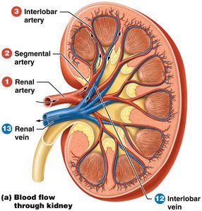
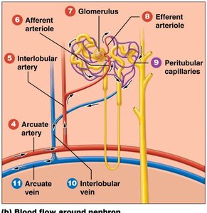
The kidneys receive 20-25% of resting cardiac output. Blood enters via the renal artery and exits via the renal vein. The renal portal system filters blood through two capillary beds: the glomerular capillaries and the peritubular capillaries.
Renal artery: Supplies blood to the kidney.
Renal vein: Drains blood from the kidney.
Glomerular capillaries: First filtration site.
Peritubular capillaries: Second filtration site, surrounding tubules.
Nephron Structure and Types
Parts of a Nephron
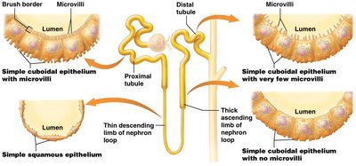
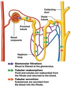
The nephron is the functional unit of the kidney, responsible for filtering blood and producing urine. It consists of:
Renal corpuscle: Glomerular capsule and glomerulus.
Renal tubule: Proximal tubule, nephron loop (descending and ascending limbs), distal tubule.
Collecting duct: Collects urine from multiple nephrons.
Renal Corpuscle
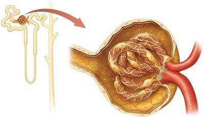
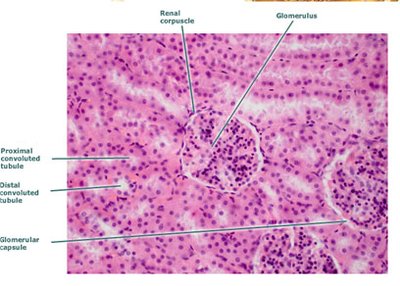
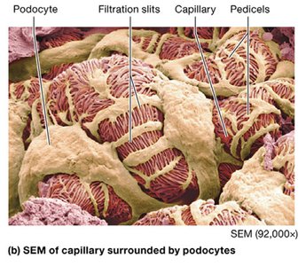
Glomerular capsule: Visceral layer (podocytes) covers capillaries; parietal layer forms outer wall.
Glomerulus: Network of capillaries where filtration occurs.
Capsular space: Area where filtered fluid collects.
Types of Nephrons
Cortical nephrons (80%): Glomerulus in outer cortex, short nephron loop.
Juxtamedullary nephrons (20%): Glomerulus in inner cortex, long nephron loop, important for urine concentration.
Renal Physiology: Urine Formation
Major Processes in Urine Formation
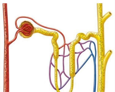
Urine formation involves three main processes:
Filtration: Removal of plasma from glomerular capillaries (≈20%).
Reabsorption: Return of 99% of water and useful solutes to blood.
Secretion: Removal of additional waste products from blood to filtrate.
Glomerular Filtration
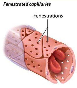
The filtration membrane permits water and small solutes to pass, but prevents plasma proteins, blood cells, and platelets from entering the filtrate. It consists of three barriers:
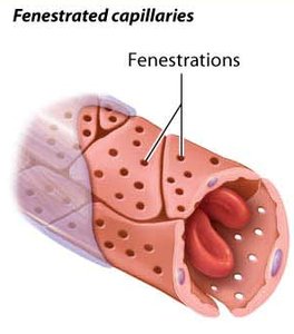
Glomerular endothelial cells: Fenestrated capillaries.
Glomerular basement membrane: Physical and charge barrier.
Filtration slits between podocytes: Final filtration barrier.
Glomerular Filtration Rate (GFR) and Net Filtration Pressure (NFP)
GFR: Amount of filtrate formed per minute by both kidneys (≈125 ml/min).
Net Filtration Pressure (NFP): Determined by hydrostatic and colloid osmotic pressures. Formula: Where GHP = glomerular hydrostatic pressure, CHP = capsular hydrostatic pressure, GCOP = glomerular colloid osmotic pressure.
Pressures favoring filtration must exceed those opposing it.
Regulation of GFR
Autoregulation: Myogenic mechanism and tubuloglomerular feedback (macula densa).
Neural control: Sympathetic nervous system.
Hormonal regulation: Renin-angiotensin-aldosterone system, ADH, ANP.
Tubular Reabsorption and Secretion
Mechanisms of Reabsorption
Passive transport: No energy required; includes osmosis and facilitated diffusion.
Active transport: Requires ATP; includes primary and secondary active transport (symporters and antiporters).
Reabsorption routes: Transcellular (through cells) and paracellular (between cells).
Symporters and Antiporters
Symporter: Moves two molecules in the same direction (e.g., Na+/glucose symporter).
Antiporter: Moves molecules in opposite directions (e.g., Na+/Ca2+ exchanger).
Reabsorption of Na+ increases reabsorption of glucose, amino acids, HCO3-, and other ions.
Reabsorption in Nephron Segments
Proximal tubule (PT): Most active segment; reabsorbs Na+, glucose, amino acids, HCO3-, water, and other ions.
Nephron loop (Loop of Henle): Descending limb reabsorbs water; ascending limb reabsorbs Na+ and Cl-.
Distal tubule (DT) and collecting duct: Early DT reabsorbs Na+ and Cl-, secretes K+ and H+; late DT and collecting duct adjust reabsorption and secretion based on body needs.
Hormonal Control of Water Reabsorption
Renin-angiotensin-aldosterone (RAA) system: Increases blood volume and pressure by increasing water reabsorption.
Antidiuretic hormone (ADH): Increases water reabsorption by increasing permeability of collecting duct.
Atrial natriuretic peptide (ANP): Decreases water and electrolyte reabsorption.
Regulation of Urine Concentration and Volume
Dilute vs. Concentrated Urine
Dilute urine: ADH not secreted; more solutes reabsorbed than water; water excreted.
Concentrated urine: ADH secreted; facultative water reabsorption; medullary osmotic gradient utilized; long nephron loops enhance concentration.
Countercurrent Mechanism
Nephron loop: Creates medullary osmotic gradient.
Vasa recta: Maintains gradient and facilitates water reabsorption.
Urine and Renal Clearance
Urinalysis and Renal Clearance
Normal urine properties: Volume (1-2L/day), pH (4.6-8), color (yellow to amber), turbidity (transparent), odor (mild).
Renal clearance: Effectiveness of kidneys at removing substances from plasma; measured by blood urea nitrogen (BUN) and plasma creatinine.
Formula for renal clearance: Where C = clearance rate, U = concentration in urine, V = urine flow rate, P = concentration in plasma.
Urine Transport, Storage, and Elimination
Ureters, Bladder, and Urethra
Ureters: Connect renal pelvis to bladder; urine moves via peristalsis and gravity.
Urinary bladder: Muscular organ posterior to pubic symphysis; stores urine.
Urethra: Drains urine from bladder to exterior; shorter in females, also serves as ejaculation duct in males.
Micturition Reflex
Stretch receptors: Stimulated when bladder volume increases (200-400ml).
Neural control: Impulses transmitted to spinal cord segments S2 and S3; detrusor muscle contracts, internal sphincter relaxes.
Voluntary control: Relaxation of external urethral sphincter required for urination.
Comparison of Male and Female Urinary Tracts
Female urethra: Exclusive passage for urine, ~4 cm long.
Male urethra: Also serves as ejaculation duct, receives sperm and fluids from accessory glands.
Summary Table: Urinary System Structures and Functions
Structure | Main Function |
|---|---|
Kidney | Filtration, hormone production, regulation of blood chemistry |
Ureter | Transport urine to bladder |
Bladder | Store urine |
Urethra | Conduct urine to exterior |
Summary Table: Nephron Segments and Functions
Segment | Main Function |
|---|---|
Renal corpuscle | Filtration |
Proximal tubule | Reabsorption of water, ions, nutrients |
Nephron loop | Reabsorption of water (DL), ions (AL) |
Distal tubule | Reabsorption and secretion |
Collecting duct | Final adjustment of urine composition |
Additional info: Academic context was added to clarify nephron function, urine formation, and regulatory mechanisms. All included images directly reinforce anatomical and physiological explanations.
