 Back
BackUrinary System and Acid-Base Balance: Structure, Function, and Regulation
Study Guide - Smart Notes
 Tailored notes based on your materials, expanded with key definitions, examples, and context.
Urinary System: Structure and Functions
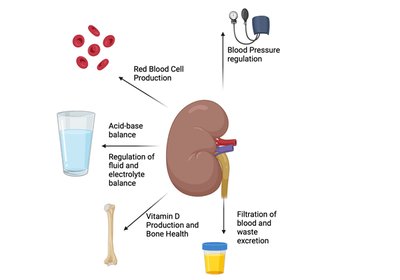
Overview of Urinary System Functions
The urinary system is essential for maintaining homeostasis by regulating the composition and volume of body fluids, blood pressure, acid-base balance, and excreting metabolic wastes. It also plays roles in hormone production and vitamin D activation.
Regulation of body water volume, blood osmolarity, and ion concentration: Kidneys filter excess water and ions, conserving them when levels are low.
Regulation of blood pressure: Kidneys control blood volume and secrete renin, initiating the renin-angiotensin-aldosterone system (RAAS).
Acid-Base balance: Kidneys generate or excrete bicarbonate and acids to maintain stable blood pH.
Excretion of metabolic waste, toxins, and drugs: Kidneys clear blood of waste products and foreign substances.
Production of erythropoietin: EPO stimulates red blood cell production in response to hypoxia.
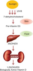
Converting vitamin D into its active form: Kidneys convert cholecalciferol to calcitriol, essential for calcium absorption.
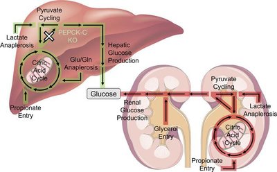
Gluconeogenesis: Kidneys produce glucose from non-carbohydrate sources during prolonged fasting.
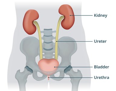
Anatomy of the Urinary System
The urinary system consists of four main structures: kidneys, ureters, urinary bladder, and urethra. Each plays a distinct role in urine formation, transport, storage, and elimination.
Kidney: Filters blood and forms urine.
Ureter: Paired tubes transporting urine from kidneys to bladder.
Urinary Bladder: Temporary storage reservoir for urine.
Urethra: Tube carrying urine from bladder to exterior.
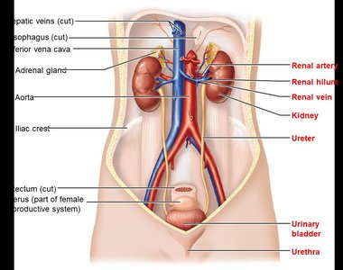
Kidney Structure and Blood Supply
Gross Anatomy and Location
The kidneys are retroperitoneal organs located on the posterior abdominal wall, typically between T12 and L3 vertebrae. The right kidney is slightly lower due to the liver's position. Each kidney is covered by supportive tissue layers and sits beneath the adrenal gland.
Renal hilum: Medial opening for ureters, blood vessels, and nerves.
Renal sinus: Internal space accessed via the hilum.
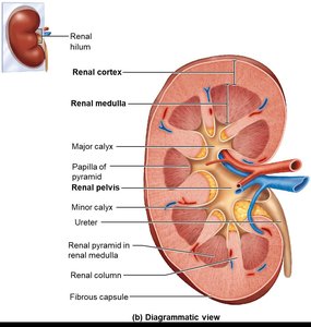
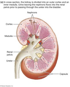
Internal Structure of the Kidney
The kidney is divided into three main regions: renal cortex, renal medulla, and renal pelvis. Each region has specialized functions in urine formation and transport.
Renal cortex: Superficial, lighter region containing most nephrons.
Renal medulla: Middle region with cone-shaped renal pyramids separated by renal columns.
Renal pelvis: Medial expansion collecting urine from calices before it enters the ureter.
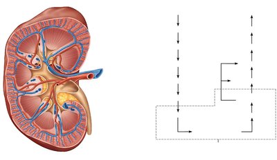
Blood Supply to the Kidney
The kidneys receive a rich blood supply, essential for filtration and homeostasis. Blood flows through a series of arteries and veins, ultimately reaching the nephron for filtration.
Renal artery: Supplies blood to the kidney.
Segmental, interlobar, arcuate, and cortical radiate arteries: Branches distributing blood within the kidney.
Renal vein: Drains filtered blood from the kidney.
Nephron: Functional Unit of the Kidney
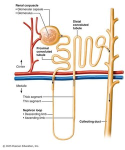
Nephron Structure
Nephrons are the structural and functional units of the kidney, responsible for filtering blood and forming urine. Each nephron consists of a renal corpuscle and renal tubule.
Renal corpuscle: Includes the glomerular (Bowman's) capsule and glomerulus, site of blood filtration.
Renal tubule: Extends from the corpuscle, consisting of proximal convoluted tubule, nephron loop (loop of Henle), and distal convoluted tubule.
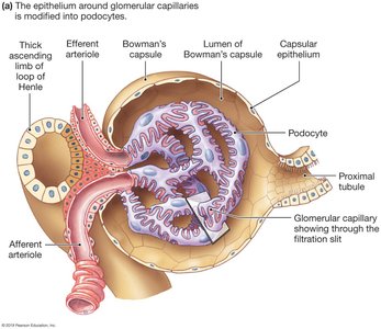
Renal Corpuscle and Filtration
The renal corpuscle is the site of initial blood filtration. The glomerulus is a ball of fenestrated capillaries, surrounded by the glomerular capsule. Podocytes cover the glomerulus, forming filtration slits.
Fenestrated capillaries: Allow passage of small solutes, but not cells or proteins.
Podocytes: Specialized cells regulating filtration.
Renal Tubule Regions
The renal tubule processes filtrate through reabsorption and secretion. Each region has distinct functions:
Proximal convoluted tubule (PCT): Major site for reabsorption of water, ions, glucose, and bicarbonate; also secretion of waste.
Nephron loop (loop of Henle): U-shaped, with descending limb permeable to water and ascending limb permeable to solutes; concentrates urine.
Distal convoluted tubule (DCT): Further reabsorption and secretion, regulated by hormones.
Collecting duct: Drains filtrate into renal pelvis; principal cells regulate water and sodium balance, intercalated cells regulate acid-base balance.
Renal Processes: Filtration, Reabsorption, and Secretion
Filtration at the Glomerulus
Filtration is a passive process driven by hydrostatic pressure, producing a cell- and protein-free filtrate. The filtration membrane consists of fenestrated endothelium, basement membrane, and podocytes.
Hydrostatic pressure in glomerular capillaries (HPGC): Promotes filtration.
Capsular space hydrostatic pressure (HPCS) and colloid osmotic pressure: Inhibit filtration.
Net filtration pressure:
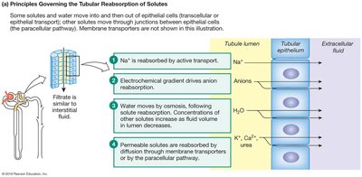
Tubular Reabsorption
Most filtered water and solutes are reabsorbed along the renal tubule. Reabsorption occurs via transcellular (through cells) or paracellular (between cells) routes, using active and passive transport mechanisms.
Na+ reabsorption: Active transport creates an electrochemical gradient for anion reabsorption.
Water reabsorption: Follows osmotic gradient generated by sodium reabsorption; aquaporins facilitate water movement.
Hormonal regulation: ADH increases water reabsorption; aldosterone increases sodium reabsorption.
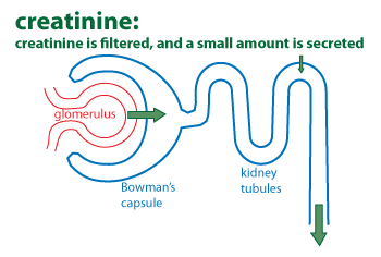
Tubular Secretion
Unwanted substances not filtered or unable to be filtered are secreted into renal tubules. This process is essential for removing excess ions, drugs, and metabolic wastes.
Substances secreted: H+, K+, NH4+, creatinine, organic acids and bases.
Example: Creatinine is filtered at the glomerulus and a small amount is secreted by the tubules.
Regulation of Kidney Function
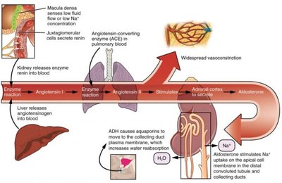
Renin-Angiotensin-Aldosterone System (RAAS)
The RAAS is a hormonal system regulating blood pressure and GFR. Renin released by the kidney initiates a cascade resulting in vasoconstriction and increased sodium and water reabsorption.
Renin: Secreted by granular cells in response to low blood pressure or NaCl.
Angiotensin II: Causes vasoconstriction and stimulates aldosterone and ADH secretion.
Aldosterone: Increases sodium reabsorption in distal tubule and collecting duct.
ADH: Increases water reabsorption by increasing aquaporins in collecting duct.
Hormonal and Homeostatic Functions of the Kidney
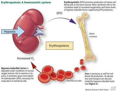
Erythropoietin Production
The kidney produces erythropoietin (EPO), a hormone that stimulates red blood cell production in the bone marrow, especially in response to hypoxia.
EPO: Promotes erythropoiesis, increasing oxygen-carrying capacity of blood.
Vitamin D Activation
The kidney converts inactive vitamin D (cholecalciferol) into its active form (calcitriol), which is essential for calcium absorption and bone health.
Calcitriol: Active form of vitamin D, produced by kidney cells containing 1-alpha-hydroxylase.
Gluconeogenesis
During prolonged fasting, kidney cells can produce glucose from non-carbohydrate sources (amino acids, lactate, glycerol) to maintain normal blood glucose levels.
Renal gluconeogenesis: Supplements hepatic glucose production, especially during starvation.
Acid-Base Balance and Renal Regulation
Chemical Buffers and Respiratory Regulation
Body fluids are maintained within a narrow pH range by chemical buffers, respiratory regulation, and renal regulation. The kidneys play a crucial role in long-term acid-base balance.
Bicarbonate buffer system: Most important in blood and extracellular fluid.
Phosphate buffer system: Effective in cells and urine.
Protein buffer system: Proteins act as amphoteric buffers.
Respiratory regulation: Lungs expel or retain CO2 to adjust blood pH.
Renal Regulation of Acid-Base Balance
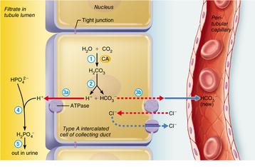
The kidneys regulate blood pH by conserving, generating, or excreting bicarbonate (HCO3-) and hydrogen ions (H+). This process is essential for compensating metabolic and respiratory acid-base disturbances.
Conservation/generation of HCO3-: Occurs in the proximal tubule and collecting duct (alpha intercalated cells).
Excretion of HCO3-: Occurs in the collecting duct (beta intercalated cells).
Ammoniagenesis: Conversion of glutamine to bicarbonate and ammonium ion, which is secreted.
Summary Table: Major Functions of the Urinary System
Function | Mechanism | Key Structure/Hormone |
|---|---|---|
Water & Ion Regulation | Filtration, reabsorption, secretion | Nephron, ADH, aldosterone |
Blood Pressure Regulation | RAAS, water volume control | Renin, angiotensin II, aldosterone |
Acid-Base Balance | Bicarbonate generation/excretion | Nephron, intercalated cells |
Waste Excretion | Filtration, secretion | Nephron, tubules |
Erythropoietin Production | Hormone secretion | Kidney, EPO |
Vitamin D Activation | Enzymatic conversion | Kidney, calcitriol |
Gluconeogenesis | Metabolic pathway | Kidney, renal cells |
