 Back
BackUrinary System: Structure and Physiology
Study Guide - Smart Notes
 Tailored notes based on your materials, expanded with key definitions, examples, and context.
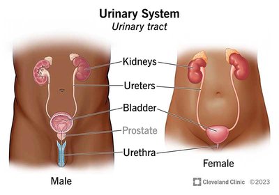
The Urinary System
Overview and Functions
The urinary system is essential for filtering waste products and excess water from the blood, producing urine, and maintaining homeostasis of pH, water, and electrolytes. The primary organs are the kidneys, ureters, bladder, and urethra.
Kidneys: Main site of blood filtration.
Ureters: Connect kidneys to bladder.
Bladder: Stores urine.
Urethra: Duct for urine expulsion.
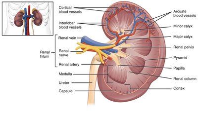
Anatomy of the Kidney
The kidney is a complex organ with distinct regions and structures that facilitate blood filtration and urine formation.
Cortex: Outer region containing nephrons.
Medulla: Inner region with renal pyramids.
Renal pelvis: Collects urine before it enters the ureter.
Blood vessels: Include renal artery, vein, and various branches for blood supply and drainage.
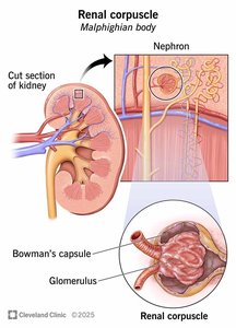
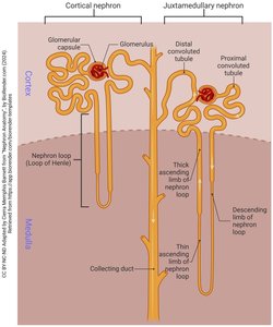
Nephron Structure and Function
Renal Corpuscle
The nephron is the functional unit of the kidney, with each kidney containing about 1 million nephrons. The renal corpuscle consists of the glomerulus (a tuft of capillaries) and Bowman’s capsule.
Glomerulus: Site of blood filtration.
Bowman's capsule: Surrounds the glomerulus and collects filtrate.
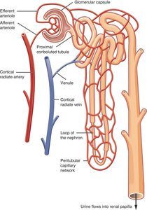
Nephron Tubules
The nephron contains several segments, each with specialized functions:
Proximal Convoluted Tubule (PCT): Reabsorbs most water, ions, glucose, and amino acids.
Loop of Henle: Descending limb reabsorbs water; ascending limb reabsorbs salt.
Distal Convoluted Tubule (DCT): Regulates Na+, K+, and water balance.
Collecting Duct: Final regulation of water and electrolytes, influenced by hormones.
Glomerular Filtration
Filtration Process
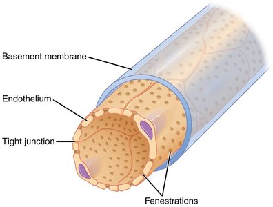
Glomerular filtration is the first step in urine formation, separating plasma from blood cells. The glomerulus is a high-pressure cluster of fenestrated capillaries, allowing water and small molecules to pass but retaining blood cells.
Fenestrated capillaries: Specialized for filtration.
Filtration slits: Created by podocytes, allow selective passage of substances.

Regulation of Filtration
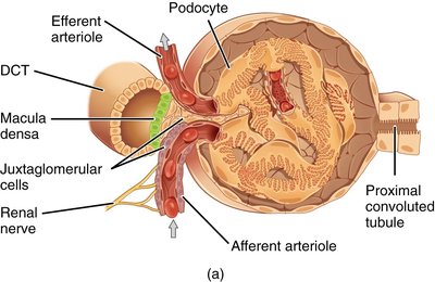
The afferent arteriole brings blood to the glomerulus, while the efferent arteriole carries it away. The juxtaglomerular apparatus (JGA) regulates blood pressure and filtration rate, with macula densa cells acting as chemoreceptors for NaCl concentration.
High NaCl: Macula densa releases ATP, causing afferent arteriole constriction and reduced GFR.
Low NaCl: Promotes dilation and increased GFR.
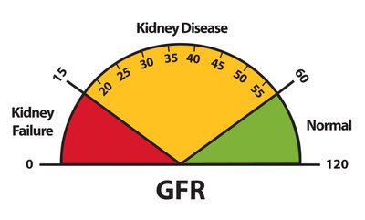
Glomerular Filtration Rate (GFR)
GFR measures the volume of filtrate produced per minute. Normal GFR is 90-120 mL/min.
99% of filtrate is reabsorbed; only 1-2L of urine produced daily.
GFR is a key indicator of kidney health.
Endocrine Regulation: RAAS System
Renin-Angiotensin-Aldosterone System (RAAS)
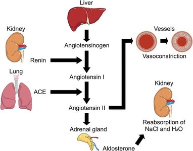
Juxtaglomerular cells sense low blood pressure and release renin, initiating the RAAS pathway.
Renin: Converts angiotensinogen (from liver) to angiotensin I.
ACE (from lungs): Converts angiotensin I to angiotensin II.
Angiotensin II: Vasoconstrictor, stimulates aldosterone release.
Aldosterone: Promotes Na+ and water reabsorption, increasing blood pressure.
ADH: Increases water reabsorption via aquaporins.
Tubular Reabsorption
Overview of Reabsorption
Tubular reabsorption is the process by which peritubular capillaries reclaim water, ions, and nutrients from the filtrate. Most reabsorption occurs in the PCT, loop of Henle, DCT, and collecting duct.
Mechanisms: Include active transport, diffusion, and osmosis.
Regulation: Hormones such as renin, ADH, and aldosterone tightly control water reabsorption.
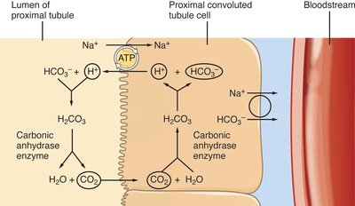
Proximal Convoluted Tubule (PCT)
The PCT recovers the bulk of filtered substances, including 60-70% of water and salts, and nearly all glucose, amino acids, and vitamins.
Bicarbonate reabsorption: Vital for pH balance, involves carbonic anhydrase and transporters.
Osmosis: Water follows solute reabsorption, moving through aquaporin channels.
Loop of Henle
The loop of Henle uses a countercurrent multiplier mechanism to concentrate urine.
Descending limb: Permeable to water, reabsorbs about 15%.
Ascending limb: Impermeable to water, actively reabsorbs ions.
Distal Convoluted Tubule (DCT) and Collecting Duct
The DCT and collecting duct fine-tune water and electrolyte recovery, influenced by aldosterone and ADH.
Principal cells: Regulate water and sodium balance.
Intercalated cells: Regulate pH by secreting protons or bicarbonate.
Summary Table: Urine Formation Steps
Step | Location | Main Function |
|---|---|---|
Glomerular Filtration | Renal Corpuscle | Filters plasma, retains cells/proteins |
Tubular Reabsorption | PCT, Loop of Henle, DCT, Collecting Duct | Reclaims water, ions, nutrients |
Tubular Secretion | PCT, DCT, Collecting Duct | Removes excess ions, wastes |
Urine Excretion | Renal pelvis, ureters, bladder, urethra | Eliminates urine from body |
Key Terms and Concepts
Nephron: Functional unit of the kidney.
Glomerular Filtration Rate (GFR): Volume of filtrate produced per minute.
Podocytes: Specialized cells forming filtration slits.
Juxtaglomerular Apparatus (JGA): Regulates blood pressure and filtration rate.
Renin-Angiotensin-Aldosterone System (RAAS): Hormonal pathway for blood pressure regulation.
Aquaporins: Water channel proteins in nephron cells.
Relevant Equations
Glomerular Filtration Rate (GFR)
GFR can be estimated using the following formula:
Bicarbonate Buffer Equation
Key reaction for pH regulation:
Practice Questions
What two structures form the renal corpuscle?
How is plasma filtered out while blood cells and large particles are retained during glomerular filtration?
What are the roles of the afferent and efferent arterioles?
What are podocytes and what is their function?
What is the function of macula densa cells and what molecule do they release to regulate filtration rate?
What are juxtaglomerular cells and what enzyme do they release when blood pressure is low?
What roles do aldosterone and antidiuretic hormone play in maintaining blood pressure?
What is the role of the proximal convoluted tubule and what molecules are filtered there?
Why is bicarbonate reabsorption in the PCT important?
What are the special protein channels used for water reabsorption?
Describe the permeability of the loop of Henle and the countercurrent mechanism.
What are the two types of specialized cells in the collecting duct and their functions?
Additional info: Academic context was added to clarify nephron structure, RAAS pathway, and mechanisms of reabsorption and filtration.
