 Back
BackUrinary System: Structure, Function, and Urine Formation
Study Guide - Smart Notes
 Tailored notes based on your materials, expanded with key definitions, examples, and context.
The Urinary System
Functions of the Urinary System
The urinary system is essential for maintaining homeostasis by removing waste products from the blood and regulating water and electrolyte balance. It also plays a role in eliminating toxins and drugs from the body.
Elimination of waste products: Includes nitrogenous wastes (such as urea, uric acid, and creatinine), toxins, and drugs.
Regulation: Maintains the balance of water, electrolytes, and acid-base in the body.
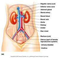
Organs of the Urinary System
The urinary system consists of four main organs, each with a specific function in the production, storage, and elimination of urine.
Kidneys: Filter blood and produce urine.
Ureters: Transport urine from the kidneys to the urinary bladder.
Urinary bladder: Stores urine until it is excreted.
Urethra: Conducts urine from the bladder to the outside of the body.
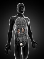
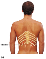
Location of the Kidneys
The kidneys are retroperitoneal organs located against the dorsal body wall, spanning the level of the T12 to L3 vertebrae. The right kidney is slightly lower than the left due to the position of the liver.
Retroperitoneal: Positioned behind the peritoneal cavity.
Protection: Partially protected by the lower ribs.
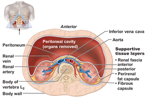
Coverings of the Kidneys
The kidneys are surrounded by three layers of supportive tissue that protect and anchor them.
Fibrous capsule: Directly surrounds each kidney, providing a tough protective layer.
Perirenal fat capsule: Cushions the kidney against mechanical injury.
Renal fascia: Outermost layer that anchors the kidney to surrounding structures.
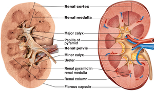
Kidney Structures
The internal anatomy of the kidney is organized into distinct regions and structures that facilitate filtration and urine collection.
Renal cortex: The outer region where filtration occurs.
Renal medulla: Contains renal pyramids, which are involved in urine concentration.
Renal pelvis: Funnel-shaped structure that collects urine and channels it to the ureter.
Renal columns: Extensions of cortex between pyramids.
Calyces: Minor and major calyces collect urine from the pyramids.
Microscopic Anatomy of the Kidney
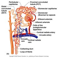
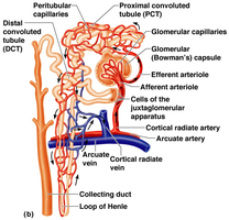
Nephron Anatomy
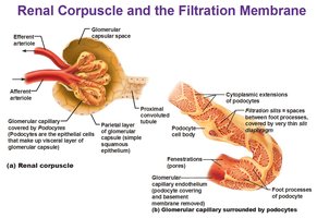
The nephron is the functional unit of the kidney, responsible for filtering blood and forming urine. Each nephron consists of a renal corpuscle and a renal tubule.
Glomerulus: A knot of capillaries where filtration begins.
Glomerular (Bowman's) capsule: Surrounds the glomerulus and collects filtrate.
Renal tubule: Includes the proximal convoluted tubule (PCT), loop of Henle, distal convoluted tubule (DCT), and ends at the collecting duct.

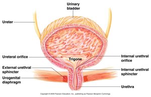
Urinary Bladder
The urinary bladder is a muscular sac that temporarily stores urine before it is excreted. It is lined with transitional epithelium to accommodate stretching.
Trigone: Triangular area formed by the openings of the ureters and urethra.
Sphincters: Internal and external sphincters control the release of urine.
Urine Formation
Processes of Urine Formation
Urine formation involves three main processes that occur in the nephron:
Glomerular filtration: Passive process where water and solutes are forced through the filtration membrane.
Tubular reabsorption: Selective process where useful substances are reabsorbed into the blood.
Tubular secretion: Additional wastes are secreted into the tubule for excretion.
Glomerular Filtration
Glomerular filtration is a nonselective, passive process driven by blood pressure. Only substances smaller than proteins pass through the filtration membrane into the glomerular capsule.
Filtration membrane: Composed of capillary endothelium, basement membrane, and podocytes.
Filtrate: Contains water, ions, glucose, amino acids, and wastes, but not proteins or blood cells.
Collecting Duct
The collecting duct receives urine from multiple nephrons and transports it through the medullary pyramids to the calyces and renal pelvis.
Function: Final concentration of urine and transport to the renal pelvis.
Characteristics of Urine
Volume and Composition
Urine is the final product of filtration, reabsorption, and secretion. Its composition reflects the body's metabolic and homeostatic needs.
Volume: 1.0 to 1.8 liters produced in 24 hours.
Filtrate vs. urine: Filtrate contains all plasma components except proteins; urine contains only wastes and unneeded substances.
Normal solutes: Sodium, potassium, urea, uric acid, creatinine, ammonia, bicarbonate ions.
Abnormal solutes: Glucose, blood proteins, red and white blood cells, hemoglobin, bile (not normally found in urine).
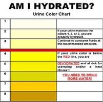
Urinalysis
Urinalysis is the examination of urine to assess its physical and chemical properties, which can indicate hydration status and health.
Color: Pale yellow to amber.
Odor: Slightly aromatic, variable.
pH: Average 6.0; range 4.5 to 8.
Specific gravity: 1.001 to 1.035.
Normal constituents: Water, urea, sodium, potassium, phosphate, sulfate ions, creatinine, uric acid, and other ions.
Microscopic Anatomy of the Ureter and Bladder
Ureter
The ureter is a muscular tube lined with transitional epithelium, facilitating the transport of urine from the kidney to the bladder.
Transitional epithelium: Allows stretching.
Muscle layers: Longitudinal and circular smooth muscle layers propel urine via peristalsis.
Bladder
The bladder wall consists of several layers, including transitional epithelium, lamina propria, and muscularis (detrusor muscle), which enable storage and expulsion of urine.
Transitional epithelium: Accommodates volume changes.
Detrusor muscle: Contracts to expel urine.
Summary Table: Urinary System Organs and Functions
Organ | Main Function |
|---|---|
Kidney | Filtration of blood, urine formation |
Ureter | Transport of urine to bladder |
Urinary bladder | Storage of urine |
Urethra | Excretion of urine |
Key Terms and Definitions
Nephron: Functional unit of the kidney responsible for urine formation.
Glomerulus: Capillary network where filtration occurs.
Bowman's capsule: Surrounds the glomerulus and collects filtrate.
Renal cortex: Outer region of the kidney.
Renal medulla: Inner region containing pyramids.
Renal pelvis: Funnel for urine collection.
Ureter: Tube transporting urine to bladder.
Urinary bladder: Muscular sac for urine storage.
Urethra: Tube for urine excretion.
Relevant Equations
Glomerular Filtration Rate (GFR):
Specific Gravity:
Additional info: Academic context was added to clarify nephron structure, urine formation processes, and urinalysis parameters for completeness.
