 Back
BackUrinary System Tracing, Disorders, and Urinalysis Study Guide
Study Guide - Smart Notes
 Tailored notes based on your materials, expanded with key definitions, examples, and context.
Q1. Trace a drop of filtrate from the glomerulus until it is excreted through the urethra.
Background
Topic: Urinary System Anatomy & Physiology
This question tests your understanding of the anatomical structures and sequence involved in the formation and excretion of urine, starting from the filtration in the nephron to its exit from the body.
Key Terms and Concepts:
Glomerulus: Capillary network where filtration begins.
Bowman's Capsule: Surrounds the glomerulus and collects filtrate.
Proximal Convoluted Tubule, Loop of Henle, Distal Convoluted Tubule: Tubular parts of the nephron where reabsorption and secretion occur.
Collecting Duct: Collects urine from multiple nephrons.
Minor Calyx, Major Calyx, Renal Pelvis: Structures that funnel urine toward the ureter.
Ureter, Urinary Bladder, Urethra: Pathways for urine excretion.
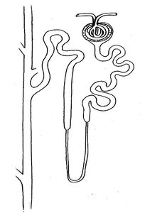
Step-by-Step Guidance
Start at the glomerulus, where blood is filtered.
The filtrate enters the Bowman's capsule.
It then flows into the proximal convoluted tubule.
Next, it travels through the loop of Henle.
After the loop, the filtrate moves into the distal convoluted tubule.
From there, it enters the collecting duct.
The collecting duct delivers the urine to the renal papilla of the renal pyramid.
Urine then passes into the minor calyx.
It continues into the major calyx.
From the major calyx, urine flows into the renal pelvis.
It then enters the ureter.
The ureter carries urine to the urinary bladder.
Finally, urine is excreted through the urethra.
Try solving on your own before revealing the answer!
Final Answer:
1. Glomerulus 2. Bowman's Capsule 3. Proximal Convoluted Tubule 4. Loop of Henle 5. Distal Convoluted Tubule 6. Collecting Duct 7. Renal Papilla (of Renal Pyramid) 8. Minor Calyx 9. Major Calyx 10. Renal Pelvis 11. Ureter 12. Urinary Bladder 13. Urethra
This sequence follows the path of filtrate as it becomes urine and is excreted from the body.
Q2. Trace an RBC through the kidney from the renal artery to the renal vein.
Background
Topic: Renal Circulation
This question assesses your knowledge of the blood flow pathway through the kidney, which is essential for understanding filtration and kidney function.
Key Terms and Concepts:
Renal Artery: Brings blood into the kidney.
Segmental, Interlobar, Arcuate, and Cortical Radiate Arteries: Branches that distribute blood within the kidney.
Afferent Arteriole: Supplies blood to the glomerulus.
Glomerulus: Site of filtration.
Efferent Arteriole: Drains blood from the glomerulus.
Peritubular Capillaries/Vasa Recta: Surround nephron tubules for exchange.
Venous return: Cortical Radiate, Arcuate, Interlobar, Renal Vein.
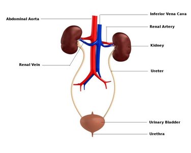
Step-by-Step Guidance
Begin at the renal artery, which enters the kidney.
Blood flows into the segmental artery.
It then moves into the interlobar artery.
Next, blood travels through the arcuate artery.
From the arcuate artery, it enters the cortical radiate artery.
Blood then passes into the afferent arteriole.
The afferent arteriole delivers blood to the glomerulus.
After filtration, blood leaves via the efferent arteriole.
It then enters the peritubular capillaries (or vasa recta, depending on nephron type).
Blood is collected by the venule.
It then flows into the cortical radiate vein.
Next, it moves into the arcuate vein.
Blood continues into the interlobar vein.
Finally, it exits the kidney through the renal vein.
Try solving on your own before revealing the answer!
Final Answer:
1. Renal Artery 2. Segmental Artery 3. Interlobar Artery 4. Arcuate Artery 5. Cortical Radiate Artery 6. Afferent Arteriole 7. Glomerulus 8. Efferent Arteriole 9. Peritubular Capillaries (or Vasa Recta) 10. Venule 11. Cortical Radiate Vein 12. Arcuate Vein 13. Interlobar Vein 14. Renal Vein
This pathway traces the flow of a red blood cell through the kidney's vascular system.
Q3. Match the urinary disorders to their causes and symptoms.
Background
Topic: Renal Pathophysiology
This question tests your ability to identify common kidney and urinary tract disorders, their causes, and associated signs and symptoms.
Key Terms:
Cystitis (lower UTI): Infection of the bladder.
Renal calculi: Kidney stones.
Pyelonephritis (upper UTI): Infection of the kidney.
Nephrosis: Disease of the glomeruli.
Polycystic Disease: Genetic disorder causing malformed renal tubules.
Diabetes Insipidus: Hyposecretion of ADH.
Step-by-Step Guidance
Read each description in the table and identify the key symptoms and causes.
Recall the definitions and typical presentations of each disorder listed in the word bank.
Match the disorder to the description based on unique features (e.g., "painful urination" for cystitis, "proteinuria" for nephrosis).
Continue matching until all disorders are paired with their correct causes and symptoms.
Try solving on your own before revealing the answer!
Final Answer:
Name of Disorder | Cause | Signs/Symptoms |
|---|---|---|
Cystitis (lower UTI) | Infection of the bladder | Painful urination, urge to urinate, cloudy urine, hematuria |
Renal calculi | Dehydration, UTI | Severe groin/flank pain, hematuria, nausea/vomiting |
Pyelonephritis (upper UTI) | Bacterial infection of the kidney | Fever, flank pain, painful urination, vomiting |
Nephrosis | Disease glomeruli | Proteinuria, body wide edema |
Polycystic Disease | Genetic disorder causing malformed renal tubules | High blood pressure, abdominal pain, hematuria, polyuria |
Diabetes Insipidus | Hyposecretion of ADH | Polydipsia, polyuria, very dilute urine, dehydration |
Each disorder is matched to its cause and characteristic symptoms.
Q4. Urinalysis: Specific Gravity, pH, and Abnormal Constituents
Background
Topic: Urinalysis and Interpretation
This question covers the normal and abnormal findings in urinalysis, including specific gravity, pH, and the significance of abnormal constituents in urine.
Key Terms and Ranges:
Specific Gravity: Normal range is 1.003–1.032.
Urine pH: Normal range is 4.5–8.0.
Abnormal Constituents: Glucose, protein, ketones, blood, bilirubin, etc.
Step-by-Step Guidance
Recall that a well-hydrated person will have urine with a lower specific gravity, while dehydration increases specific gravity.
Remember the normal pH range for urine and how diet can affect it (vegetable-rich diets make urine more alkaline, protein-rich diets make it more acidic).
Review the abnormal constituents of urine and match them to their associated disorders and causes (e.g., ketonuria with diabetes mellitus or starvation).
Use the provided table to match each abnormal finding to its possible cause and related disorder.
Try solving on your own before revealing the answer!
Final Answer:
Specific Gravity: Well-hydrated person ≈ 1.003; Dehydrated person ≈ 1.032
Urine pH: Range is 4.5–8.0; Vegetable-rich diet = more alkaline; Protein-rich diet = more acidic
Abnormal Constituents and Disorders:
Pyuria: Inflammation of urinary tract
Nitrites: Bacterial infection of urinary tract
Urobilinogenuria: Liver disease
Albuminuria: Excessive exertion, pregnancy, heart failure, severe hypertension, glomerulonephritis
Hematuria: Bleeding urinary tract, glomerulonephritis
Hemoglobinuria: Transfusion reaction, hemolytic anemia, severe burns
Ketonuria: Diabetes mellitus, starvation, low-carb diets
Bilirubinuria: Liver disease, bile duct obstruction
Glycosuria: Excess sugar intake, diabetes mellitus
Normal urine: yellow, clear, non-foamy; Abnormal: red/brown/amber (hematuria, bilirubinuria), cloudy (UTI, crystals), foamy (albuminuria)
These findings help diagnose hydration status, dietary effects, and various renal or systemic disorders.
