 Back
BackAmino Acids: Structure, Classification, and Biochemical Roles
Study Guide - Smart Notes
 Tailored notes based on your materials, expanded with key definitions, examples, and context.
Amino Acids
Definition and Biological Importance
Amino acids are organic compounds that serve as the building blocks of proteins. They play crucial roles as precursors for nucleic acids, serve as energy sources, and participate in various metabolic pathways.
Protein constituents: Amino acids are linked by peptide bonds to form proteins.
Precursors: They are precursors for nucleic acids and other biomolecules.
Energy source: Amino acids can be catabolized to provide energy.
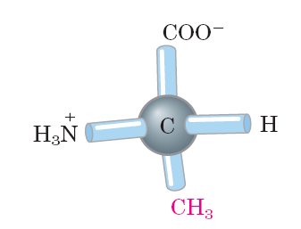
General Structure of α-Amino Acids
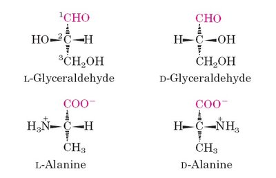
All standard amino acids (except glycine) have a central (α) carbon atom bonded to four different groups: an amino group (–NH2), a carboxyl group (–COOH), a hydrogen atom, and a variable side chain (R group) that determines the amino acid's identity.
Chirality: The α-carbon is a chiral center (except in glycine), resulting in optical isomers (L and D forms).
Biological proteins: Only L-amino acids are found in proteins.
Zwitterions and Acid-Base Properties
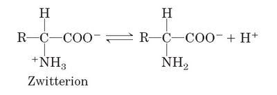
At physiological pH (~7.4), amino acids exist as zwitterions, molecules with both positive (amino group) and negative (carboxyl group) charges but a net charge of zero.
Zwitterion formation: The amino group is protonated (–NH3+), and the carboxyl group is deprotonated (–COO−).
Amphoteric nature: Amino acids can act as both acids and bases (ampholytes).
Classification of Amino Acids
Based on R Group Properties
Amino acids are classified according to the chemical nature of their side chains (R groups):
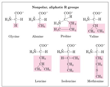
1. Nonpolar, Aliphatic R Groups
Hydrophobic side chains, often found in the interior of proteins.
Examples: Glycine, Alanine, Proline, Valine, Leucine, Isoleucine, Methionine.
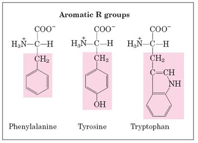
2. Aromatic R Groups
Contain aromatic rings; can participate in hydrophobic interactions and absorb UV light.
Examples: Phenylalanine, Tyrosine, Tryptophan.
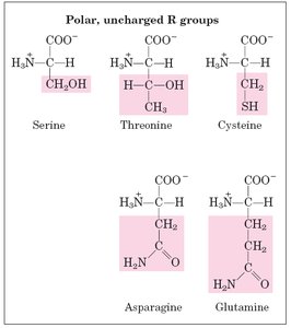
3. Polar, Uncharged R Groups
Side chains can form hydrogen bonds; often found on protein surfaces.
Examples: Serine, Threonine, Cysteine, Asparagine, Glutamine.
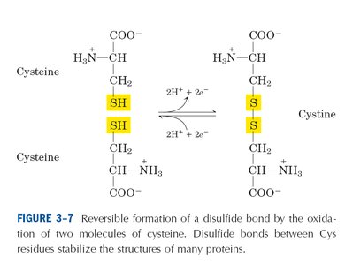
4. Sulfur-Containing Amino Acids
Cysteine can form disulfide bonds (cystine) important for protein structure.
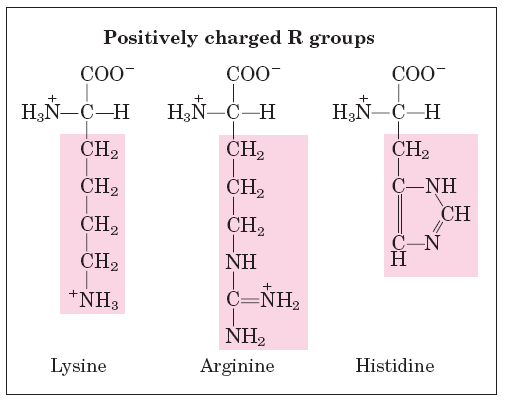
5. Positively Charged (Basic) R Groups
Side chains are positively charged at physiological pH.
Examples: Lysine, Arginine, Histidine.
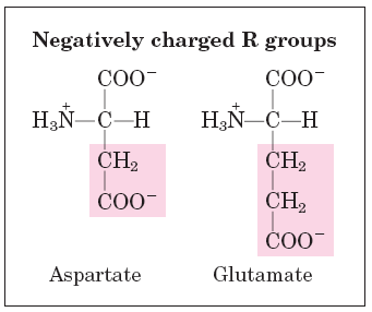
6. Negatively Charged (Acidic) R Groups
Side chains are negatively charged at physiological pH.
Examples: Aspartate, Glutamate.
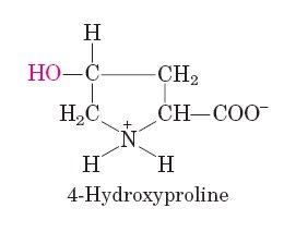
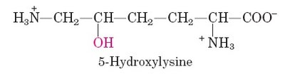
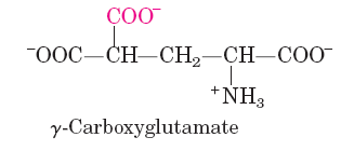
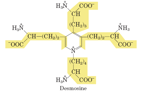
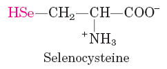
Special Amino Acids
Some amino acids are found only in specific proteins or as post-translational modifications (e.g., hydroxyproline in collagen, selenocysteine in certain enzymes).
Acid-Base Properties and Titration Curves
Ionization and Buffering
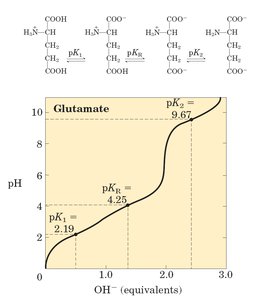
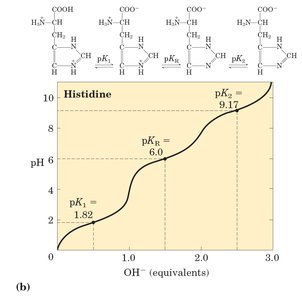
Amino acids have at least two ionizable groups (α-amino and α-carboxyl), and some have ionizable side chains. Their ionization state depends on pH, which affects their net charge and buffering capacity.
Titration curve: Shows the change in charge as pH increases.
Isoelectric point (pI): The pH at which the amino acid has no net charge.
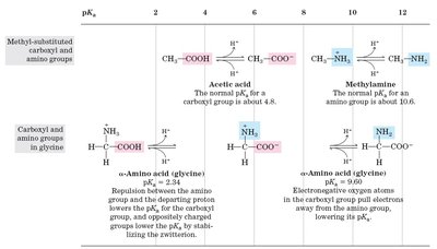
Effect of R Group Ionization
Amino acids with ionizable side chains (e.g., glutamate, histidine) have more complex titration curves and additional buffering regions.
Summary Table: Properties of Common Amino Acids
Amino acid | Abbreviation | pKa (COOH) | pKa (NH3+) | pKa (R group) | pI | Hydropathy index |
|---|---|---|---|---|---|---|
Glycine | Gly, G | 2.34 | 9.60 | - | 5.97 | -0.4 |
Alanine | Ala, A | 2.34 | 9.69 | - | 6.01 | 1.8 |
Valine | Val, V | 2.32 | 9.62 | - | 5.96 | 4.2 |
Glutamate | Glu, E | 2.19 | 9.67 | 4.25 | 3.22 | -3.5 |
Lysine | Lys, K | 2.18 | 8.95 | 10.53 | 9.74 | -3.9 |
Peptide Bond and Protein Structure
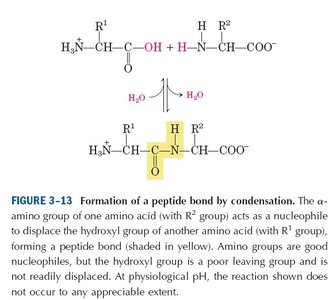
Peptide Bond Formation
Amino acids are linked by peptide bonds, formed by condensation (removal of water) between the α-carboxyl group of one amino acid and the α-amino group of another.
Peptide bond: A covalent bond with partial double-bond character, restricting rotation.
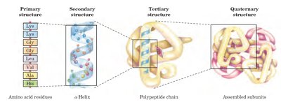
Levels of Protein Structure
Primary structure: Linear sequence of amino acids.
Secondary structure: Local folding (e.g., α-helix, β-sheet) stabilized by hydrogen bonds.
Tertiary structure: Three-dimensional folding of a single polypeptide chain.
Quaternary structure: Assembly of multiple polypeptide subunits.
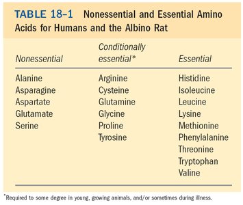
Essential and Non-Essential Amino Acids
Classification Based on Dietary Requirement
Essential amino acids: Cannot be synthesized by the body and must be obtained from the diet (e.g., histidine, isoleucine, leucine, lysine, methionine, phenylalanine, threonine, tryptophan, valine).
Non-essential amino acids: Can be synthesized by the body (e.g., alanine, asparagine, aspartate, glutamate, serine).
Conditionally essential: Required in the diet under certain conditions (e.g., arginine, cysteine, glutamine, glycine, proline, tyrosine).
Biosynthesis and Catabolism of Amino Acids
Biosynthesis
Non-essential amino acids are synthesized via simple metabolic pathways, while essential amino acids require more complex pathways. Glutamate and glutamine play central roles as amino group donors in amino acid biosynthesis.
Glutamate: Entry point for NH4+ into amino acid metabolism.
Glutamine: Formed from glutamate by glutamine synthetase; serves as an amino group donor.
Catabolism
Amino acid catabolism involves removal of the amino group (transamination and deamination), followed by oxidation of the carbon skeleton via the citric acid cycle. The amino group is ultimately converted to urea for excretion.
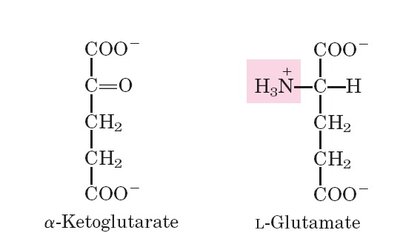
Transamination: Transfer of amino group to α-ketoglutarate, forming glutamate.
Deamination: Removal of amino group from glutamate by glutamate dehydrogenase, releasing NH4+.
Urea cycle: Conversion of toxic ammonia to urea in the liver.
Summary
Amino acids are fundamental to protein structure and function.
Their classification is based on the properties of their side chains.
They exhibit unique acid-base behavior and serve as metabolic intermediates.
Understanding their biosynthesis and catabolism is essential for grasping metabolic regulation and human nutrition.
