 Back
BackCh 11: Biological Membranes and Transport: Structure, Dynamics, and Function
Study Guide - Smart Notes
 Tailored notes based on your materials, expanded with key definitions, examples, and context.
Biological Membranes and Transport
Introduction
Biological membranes are essential components of all living cells, providing structural integrity, compartmentalization, and regulation of molecular traffic. This chapter explores the composition, architecture, dynamics, and transport mechanisms associated with biological membranes.
The Composition and Architecture of Membranes
Lipid Aggregates: Micelles, Bilayers, and Vesicles
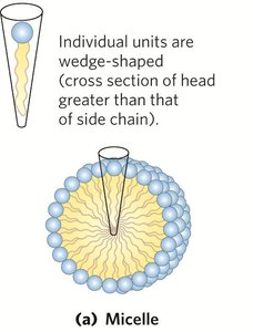
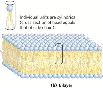
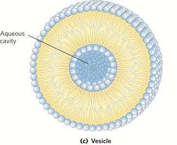
Membrane lipids are amphipathic molecules that spontaneously form organized structures in aqueous environments due to hydrophobic interactions. The three principal types of lipid aggregates are micelles, bilayers, and vesicles.
Micelles: Spherical structures formed by amphipathic molecules with hydrophobic tails inward and hydrophilic heads outward. Favored when the head group is larger than the acyl chain cross-section.
Bilayers: Two lipid monolayers (leaflets) form a two-dimensional sheet. Favored when the head group and acyl chain cross-sections are similar.
Vesicles (Liposomes): Spherical bilayers enclosing an aqueous cavity, formed when a bilayer sheet folds back on itself.
Fluid Mosaic Model and Membrane Function
The fluid mosaic model describes membranes as dynamic structures with proteins and lipids capable of lateral movement. This architecture allows for selective permeability, cell signaling, and adaptability to environmental changes.
Membranes permit shape changes, exocytosis, endocytosis, and cell division.
Proteins serve as transporters, receptors, ion channels, and adhesion molecules.
Endomembrane System and Membrane Trafficking
The endomembrane system includes organelles surrounded by single or double membranes, such as the ER, Golgi apparatus, lysosomes, nucleus, mitochondria, and chloroplasts. Membrane trafficking involves the movement and modification of lipids and proteins between these compartments.
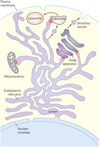
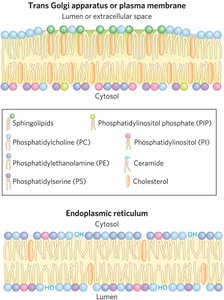
Membrane Lipid Asymmetry
Lipids are asymmetrically distributed between the two leaflets of the plasma membrane, with sphingolipids and cholesterol often replacing phosphatidylcholine in the outer leaflet during trafficking.
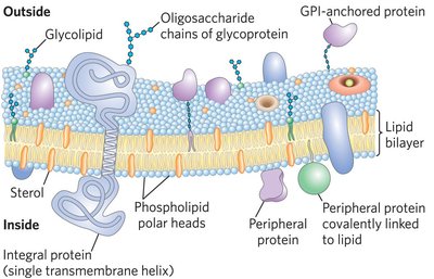
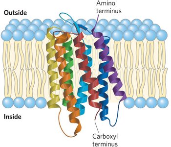
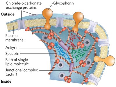
Membrane Proteins: Types and Topology
Classification of Membrane Proteins
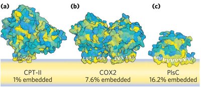
Integral membrane proteins: Firmly embedded within the lipid bilayer, often spanning the membrane.
Peripheral membrane proteins: Associate with the membrane via electrostatic interactions and hydrogen bonds.
Amphitropic proteins: Reversibly associate with membranes, found in both membranes and cytosol.
Monotopic proteins: Interact with only one leaflet of the membrane.
Bitopic proteins: Span the bilayer once with a single hydrophobic sequence.
Polytopic proteins: Cross the membrane multiple times, typically as α-helices.
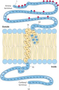
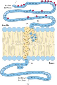
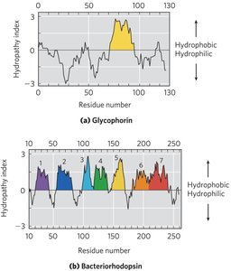
Membrane Protein Topology and Prediction
Integral membrane protein topology can often be predicted from the amino acid sequence. An α-helical segment of 20–25 residues is sufficient to span the bilayer. Hydropathy plots are used to identify hydrophobic transmembrane segments.
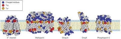
β-Barrel Proteins and Amino Acid Distribution
Some membrane proteins form β-barrels, with alternating hydrophobic and hydrophilic residues. Aromatic residues (Tyr, Trp) often anchor proteins at the membrane interface, and positively charged residues (Lys, Arg) are more common on the cytoplasmic face (positive-inside rule).
Membrane Dynamics
Lipid Mobility and Membrane Fluidity
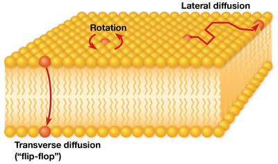
Lipids in membranes exhibit various types of motion, contributing to membrane fluidity:
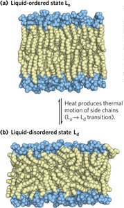
Liquid-ordered (Lo) state: Gel-like, with constrained motion.
Liquid-disordered (Ld) state: High lateral and rotational mobility.
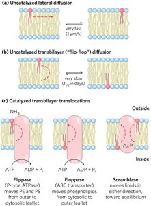
Lateral and Transverse Diffusion
Lipids can rotate and diffuse laterally within a leaflet rapidly. Transverse (flip-flop) diffusion is rare and slow but can be catalyzed by specific enzymes.
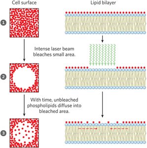
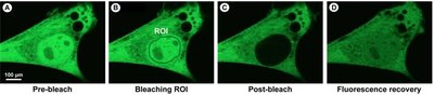
Experimental Measurement of Lipid Mobility
Fluorescence recovery after photobleaching (FRAP) is used to measure the rate of lateral diffusion of lipids and proteins in membranes.
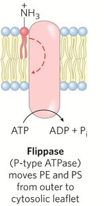
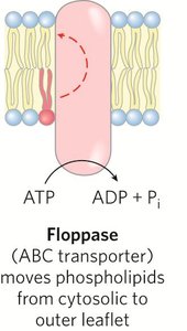
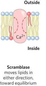
Enzymatic Catalysis of Lipid Translocation
Flippases: Move phosphatidylethanolamine (PE) and phosphatidylserine (PS) from the outer to the cytoplasmic leaflet (ATP-dependent).
Floppases: Move phospholipids and sterols from the cytoplasmic to the outer leaflet (ATP-dependent).
Scramblases: Move lipids in either direction toward equilibrium (not ATP-dependent; some require Ca2+).
Hop Diffusion and Membrane Microdomains
Lipid molecules diffuse laterally within small regions, with occasional 'hops' between regions. Microdomains (rafts) enriched in cholesterol and sphingolipids are thicker and more ordered, influencing protein localization and signaling.
Solute Transport Across Membranes
Types of Transport
Simple diffusion: Nonpolar compounds cross membranes unassisted down their concentration gradient.
Facilitated diffusion (passive transport): Polar compounds and ions require specific protein carriers to move down their gradient.
Active transport: Movement against a concentration or electrochemical gradient, requiring energy input.
Membrane Potential and Electrochemical Gradient
The membrane potential (Vm) is the electrical gradient across a membrane, influencing ion movement. The electrochemical gradient combines chemical and electrical gradients to determine solute movement direction.
Transport Proteins: Transporters and Ion Channels
Transporters: Bind solutes and undergo conformational changes to move them across membranes; can be passive or active.
Ion channels: Provide aqueous pores for rapid ion movement; typically gated and selective, not saturable.
Passive Transport: Glucose Transporters
GLUT1 mediates passive glucose transport in erythrocytes, following Michaelis-Menten-like kinetics:
where is the initial velocity, is the external glucose concentration, and is the transport constant.
Active Transport: P-Type, V-Type, F-Type ATPases, and ABC Transporters
P-type ATPases: Undergo phosphorylation during the transport cycle (e.g., Na+/K+ ATPase, Ca2+ ATPase).
V-type ATPases: Acidify intracellular compartments by pumping protons.
F-type ATPases (ATP synthases): Synthesize ATP using a proton gradient.
ABC transporters: Use ATP hydrolysis to transport a wide variety of substrates, including drugs and lipids.
Secondary Active Transport
Secondary active transport couples the uphill movement of one solute to the downhill movement of another, such as the Na+-glucose symporter in intestinal cells.
Equations for Free-Energy Change in Transport
Uncharged solute:
Ion (charged solute): where is the ion charge, is the Faraday constant, and is the membrane potential.
Aquaporins and Ion Channels
Aquaporins facilitate rapid water movement across membranes, while ion-selective channels allow specific ions to cross in response to voltage or ligand binding. These channels are critical for processes such as nerve impulse conduction and osmoregulation.
Summary Table: Major Membrane Transporters
Transporter Type | Energy Source | Direction | Example |
|---|---|---|---|
Simple diffusion | None | Down gradient | O2, CO2 |
Facilitated diffusion | None | Down gradient | GLUT1 (glucose) |
Primary active transport | ATP hydrolysis | Against gradient | Na+/K+ ATPase |
Secondary active transport | Ion gradient | Against gradient | Na+-glucose symporter |
Ion channel | None | Down gradient | K+ channel |
Aquaporin | None | Down gradient | AQP1 |
Additional info: This summary integrates foundational concepts from the Lehninger Principles of Biochemistry and expands on the provided lecture content for comprehensive exam preparation.
