 Back
BackCell Structure and Microscopy: Foundations for Biochemistry
Study Guide - Smart Notes
 Tailored notes based on your materials, expanded with key definitions, examples, and context.
A2.2 Cell Structure
Introduction to Cell Theory
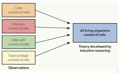
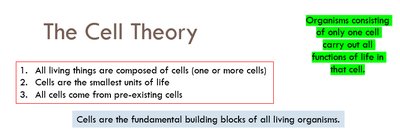
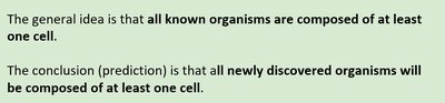
Cell theory is a fundamental concept in biology and biochemistry, stating that all living organisms are composed of cells, which are the basic structural and functional units of life. This theory was developed through inductive reasoning based on multiple observations of cellular structures in various tissues.
Key Point 1: All living things are composed of cells (one or more).
Key Point 2: Cells are the smallest units of life.
Key Point 3: All cells come from pre-existing cells.
Example: Cork, embryos, elder pith, and toad cartilage all consist of cells, supporting the cell theory.
Features Common to All Cells
All cells share certain structural features essential for life. These include the presence of DNA as genetic material, a cytoplasm primarily composed of water, and a plasma membrane made of lipids.
Cytoplasm: The site of metabolic activity; water is the main component.
Plasma Membrane: Controls entry and exit of substances; composed of lipids.
DNA: Genetic material; in eukaryotes, DNA is bound in a nucleus with histone proteins, while in prokaryotes, it is free in the cytoplasm.
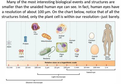
A2.2.2 Microscopy Skills
Microscope Structure and Function
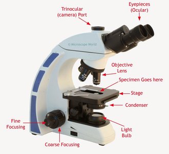
Microscopy is a key technique for investigating cell structure. Understanding the parts and operation of a microscope is essential for biochemistry students.
Eyepieces (Ocular): Used to view the specimen.
Objective Lens: Magnifies the specimen.
Stage: Holds the slide.
Condenser: Focuses light onto the specimen.
Coarse and Fine Focusing: Adjusts clarity and focus.
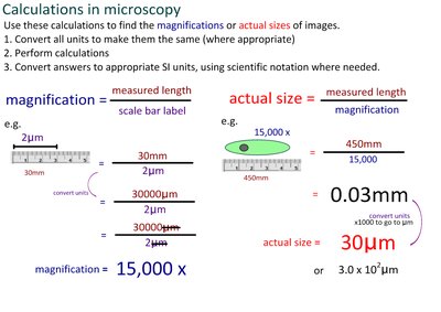
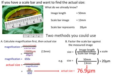
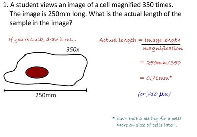
Calculating Magnification and Specimen Size
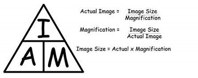
Magnification and actual size calculations are fundamental skills in microscopy. The formulas are:
Magnification:
Actual Size:
Image Size:
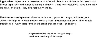
Microscopy Techniques
Modern microscopy includes advanced techniques such as electron microscopy, freeze fracture, cryogenic electron microscopy, and fluorescent staining. These methods provide higher resolution and allow visualization of cellular structures and biomolecules.
Electron Microscopy: Offers much higher resolution and magnification than light microscopy.
Freeze Fracture: Used to study internal surfaces of cells, especially membranes.
Cryogenic Electron Microscopy: Allows visualization of proteins and biomolecules that do not crystallize easily.
Fluorescent Stains: Used to highlight specific cell structures, often with antibodies.
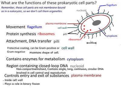
A2.2.4 Prokaryote and Eukaryote Cell Structure
Prokaryote Cell Structure
Prokaryotes, such as Gram-positive eubacteria, have a simpler cell structure compared to eukaryotes. Key components include:
Cell Wall: Provides structural support.
Plasma Membrane: Controls entry and exit of substances.
Cytoplasm: Site of metabolic activity.
Naked DNA: Circular DNA free in the cytoplasm.
70S Ribosomes: Site of protein synthesis.
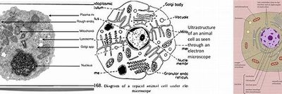
Eukaryote Cell Structure
Eukaryotic cells are compartmentalized and contain membrane-bound organelles. Common features include:
Plasma Membrane: Encloses the cytoplasm.
Nucleus: Contains chromosomes made of DNA bound to histones, surrounded by a double membrane with pores.
80S Ribosomes: Larger ribosomes for protein synthesis.
Organelles: Mitochondria, endoplasmic reticulum, Golgi apparatus, lysosomes, vacuoles, and vesicles.
Cytoskeleton: Microtubules and microfilaments provide structural support.
A2.2.12 Origin of Eukaryotic Cells by Endosymbiosis
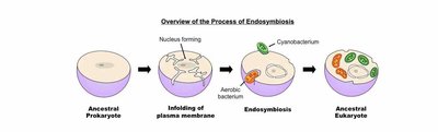
Endosymbiotic Theory
The endosymbiotic theory explains the origin of mitochondria and chloroplasts in eukaryotic cells. It proposes that these organelles originated from symbiotic relationships between ancestral prokaryotes.
Key Evidence: Presence of 70S ribosomes, naked circular DNA, and the ability to replicate in mitochondria and chloroplasts.
Process: Ancestral prokaryote engulfed aerobic bacteria (mitochondria) and cyanobacteria (chloroplasts), leading to modern eukaryotes.
A2.2.13 Cell Differentiation and Multicellularity
Cell Differentiation
Cell differentiation is the process by which cells develop specialized functions in multicellular organisms. This is achieved through selective gene expression, often triggered by environmental changes.
Stem Cells: Undifferentiated cells capable of becoming any cell type.
Gene Expression: Determines cell function by activating or deactivating specific genes.
Example: Nerve cells, muscle cells, and blood cells are specialized through differentiation.
Evolution of Multicellularity
Multicellularity has evolved independently in various lineages, allowing for larger body size, cell specialization, and increased complexity. Most biomass on Earth is unicellular, but multicellular organisms dominate in plants and animals.
Advantages: Longer lifespans, ability to exploit new niches, and increased complexity.
Summary Table: Prokaryote vs. Eukaryote Cell Structure
Feature | Prokaryote | Eukaryote |
|---|---|---|
Size | ~10 microns | ~100 microns |
Cell Wall | Present | Present in plants/fungi |
DNA | Naked, circular, free in cytoplasm | Bound to histones, linear, in nucleus |
Ribosomes | 70S | 80S |
Organelles | None | Membrane-bound |
Additional info:
These notes expand on the original content by providing definitions, formulas, and examples relevant to biochemistry students. The included images directly reinforce the explanations of cell theory, microscopy, cell structure, and endosymbiosis.
