 Back
Chapter 4: Nucleic Acids – Structure, Properties, and Biological Functions
Study Guide - Smart Notes
 Tailored notes based on your materials, expanded with key definitions, examples, and context.
Nucleic Acids: Structure and Nomenclature
Bases, Nucleosides, and Nucleotides
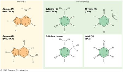
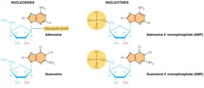
Nucleic acids are polymers composed of nucleotide monomers, each consisting of a nitrogenous base, a pentose sugar, and a phosphate group. The nomenclature distinguishes between bases, nucleosides (base + sugar), and nucleotides (base + sugar + phosphate).
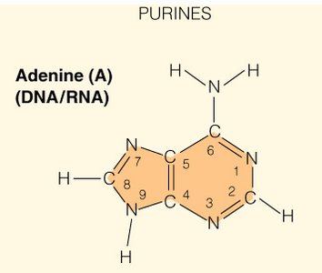
Bases: Purines (adenine, guanine) and pyrimidines (cytosine, thymine, uracil).
Nucleosides: Adenosine, guanosine, cytidine, uridine, thymidine.
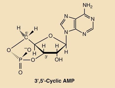
Nucleotides: Adenosine 5'-monophosphate (AMP), Guanosine 5'-monophosphate (GMP), etc.
Example: Adenine is a purine base; adenosine is the corresponding nucleoside; AMP is the nucleotide.
Tautomeric Forms and Ionization
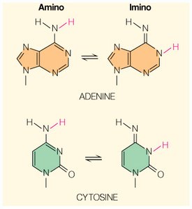
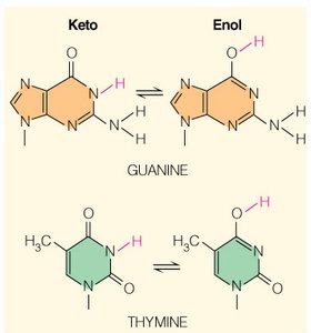
Nucleic acid bases can exist in different tautomeric forms (amino/imino, keto/enol), which influence hydrogen bonding and base pairing. Ionization states are determined by pKa values and physiological pH.
Tautomerism: Amino and imino forms for adenine and cytosine; keto and enol forms for guanine and thymine.
Ionization: At physiological pH (6.5–8.5), bases are generally uncharged.
Phosphate | Base | pKa1 | pKa2 | pKa | Reaction (Loss of Proton from) |
|---|---|---|---|---|---|
5' AMP | Adenine | 0.9 | 6.1 | 3.8 | N-1 |
5' GMP | Guanine | 0.8 | 6.1 | 2.4 | N-1 |
5' UMP | Uracil | 1.0 | 6.4 | 9.5 | N-3 |
5' CMP | Cytosine | 0.8 | 6.3 | 4.5 | N-3 |
DNA and RNA: Structural Differences and Biological Significance
Chemical Differences Between DNA and RNA
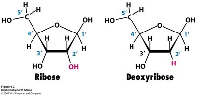
DNA and RNA differ in their sugar component and one of their bases. DNA contains deoxyribose and thymine, while RNA contains ribose and uracil. The absence of the 2'-OH group in DNA makes it more stable than RNA.
DNA: Deoxyribose sugar, bases A, T, G, C.
RNA: Ribose sugar, bases A, U, G, C.
Stability: DNA is more stable due to lack of 2'-OH; RNA is more susceptible to hydrolysis.
Base Pairing and Chargaff’s Rules
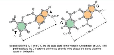
Base pairing in DNA follows the Watson-Crick model: A pairs with T, and G pairs with C. Chargaff’s rules state that the mole percent of A equals T, and G equals C in double-stranded DNA.
Base Pairing: A–T and G–C pairs are stabilized by hydrogen bonds.
Distance: The C1' carbons on the two strands are 1.08 nm apart for both pairs.
Nucleotide Functions
Biological Roles of Nucleotides
Nucleotides serve multiple functions in cells, including energy transfer, signaling, and as coenzyme components.
Energy: ATP is the primary energy carrier; GTP, CTP, and UTP are used in protein, lipid, and carbohydrate synthesis, respectively.
Signaling: cAMP and cGMP act as second messengers in signal transduction.
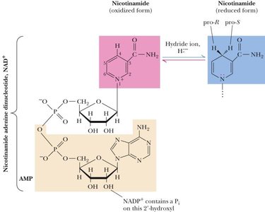
Coenzymes: NAD+, NADP+, FMN, and FAD are essential for redox reactions.
UV Absorbance Features of DNA and Proteins
UV-Vis Absorbance and Beer-Lambert Law
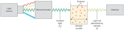
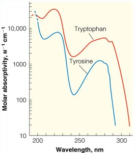
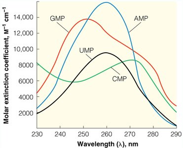
Nucleic acids and proteins absorb ultraviolet light, but at different wavelengths. The Beer-Lambert Law relates absorbance to concentration, allowing quantification of DNA and RNA.
DNA: Maximum absorbance at 260 nm; A260/A280 ratio of 1.8–2.0 indicates pure DNA.
Protein: Maximum absorbance at 280 nm; A260/A280 ratio of ~0.5 indicates pure protein.
Beer-Lambert Law:
DNA Structure: Primary, Secondary, and Tertiary
Levels of Nucleic Acid Structure
Nucleic acids exhibit hierarchical structural organization:
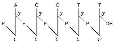
Primary: Linear sequence of nucleotides (5' to 3' direction).
Secondary: Local folding, such as the double helix.
Tertiary: Higher-order folding, including supercoiling and cruciforms.
DNA Double Helix and Stabilizing Forces
The DNA double helix is stabilized by hydrogen bonds between base pairs and base stacking interactions. The helix has ~10.4 base pairs per turn, and the rise per base pair is 0.34 nm.
Hydrogen Bonds: 2–6 kJ/mol per bond.
Base Stacking: 4 kJ/mol per base pair.
Helical Parameters: 36° angle between stacked base pairs; 10 bp/turn.
DNA Topology and Supercoiling
Supercoiling and Linking Number
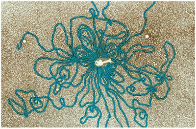
DNA topology is described by the linking number (L), twist (T), and writhe (W). Supercoiling is a tertiary structural feature important for DNA compaction and regulation.
Linking Number (L): Number of times one strand crosses the other; only changes if DNA is broken.
Twist (T): Number of helical turns.
Writhe (W): Number of superhelical turns.
Equation:
DNA Denaturation and Renaturation
Helix-Coil Transition and Hyperchromic Effect
DNA denaturation involves the separation of double-stranded DNA into single strands, promoted by heat, alkali, or electrostatic repulsion. The process is reversible, and renaturation occurs upon slow cooling.
Hyperchromic Effect: Denatured DNA absorbs more UV light than native DNA.
Cooperative Transition: Melting occurs over a narrow temperature range.
Palindromic DNA, Restriction Enzymes, and Molecular Cloning
Palindromic Sequences and Restriction Enzymes
Palindromic DNA sequences are symmetrical and often serve as recognition sites for restriction enzymes, which are essential tools in molecular cloning.
Restriction Enzymes: Over 3000 identified; more than 600 commercially available.
Recognition Sites: Typically 4–8 base pairs, often palindromic.
Summary Table: Nucleic Acid Bases
Base | Nucleoside | Nucleotide |
|---|---|---|
Adenine | Adenosine | Adenosine 5'-monophosphate |
Guanine | Guanosine | Guanosine 5'-monophosphate |
Cytosine | Cytidine | Cytidine 5'-monophosphate |
Uracil | Uridine | Uridine 5'-monophosphate |
Thymine | Thymidine | Deoxythymidine 5'-monophosphate |
Additional info:
DNA is the carrier of genetic information, as demonstrated by classic experiments (Avery, Hershey & Chase).
DNA replication is semiconservative, with each strand serving as a template for a new strand.
Electrophoresis and agarose gels are used to analyze DNA topology and supercoiling.
