 Back
DNA Replication, Transcription, and Translation: Concepts and Connections
Study Guide - Smart Notes
 Tailored notes based on your materials, expanded with key definitions, examples, and context.
DNA Replication
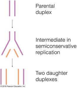
Semiconservative Replication
DNA replication is the process by which a cell duplicates its genome prior to cell division. The semiconservative model states that each daughter DNA molecule consists of one parental strand and one newly synthesized strand.
Parental duplex: The original double-stranded DNA.
Intermediate: During replication, the two strands separate and serve as templates.
Daughter duplexes: Each new DNA molecule contains one old and one new strand.
Replication Fork and Enzymes
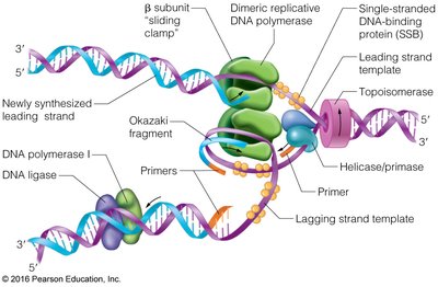
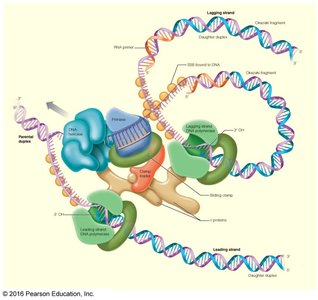
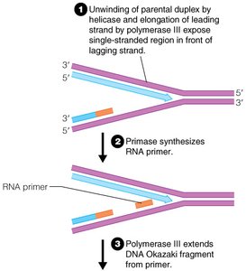
Replication begins at origins and proceeds bidirectionally. The replication fork is the site where DNA strands are separated and new DNA is synthesized.
Helicase: Unwinds the DNA double helix.
Primase: Synthesizes RNA primers for DNA polymerase.
Single-stranded binding protein (SSB): Stabilizes separated strands.
DNA polymerase III: Main enzyme for DNA synthesis.
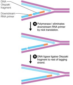
DNA polymerase I: Removes RNA primers and fills gaps.
DNA ligase: Joins Okazaki fragments on the lagging strand.
Topoisomerase: Relieves supercoiling tension.
Leading and Lagging Strand Synthesis
DNA polymerase synthesizes DNA in the 5' to 3' direction. The leading strand is synthesized continuously, while the lagging strand is synthesized discontinuously in short fragments called Okazaki fragments.
Okazaki fragments: Short DNA segments synthesized on the lagging strand.
DNA ligase: Joins Okazaki fragments to form a continuous strand.
DNA Polymerase Reaction Mechanism
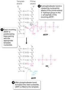
DNA polymerase catalyzes the formation of phosphodiester bonds between nucleotides. The 3'-OH group of the primer attacks the α-phosphate of the incoming dNTP, releasing pyrophosphate.
Phosphodiester bond: Covalent bond joining nucleotides.
Pyrophosphate (PPi): Released as a byproduct.
Equation:
Linear Chromosome Replication and Telomeres
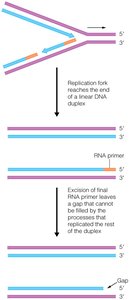
Linear chromosomes face the end-replication problem, where the removal of the final RNA primer leaves a gap at the 5' end. Eukaryotes solve this with telomeres and the enzyme telomerase.
Telomeres: Repetitive DNA sequences at chromosome ends.
Telomerase: Extends telomeres using an RNA template.
Experimental Methods: PCR and Sanger Sequencing
Polymerase Chain Reaction (PCR)
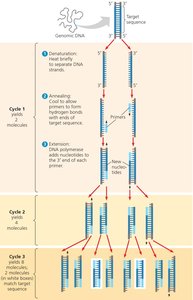
PCR is a technique used to amplify specific DNA sequences. It involves repeated cycles of denaturation, annealing, and extension.
Denaturation: Heating separates DNA strands (98°C).
Annealing: Cooling allows primers to bind (55°C).
Extension: DNA polymerase synthesizes new DNA (72°C).
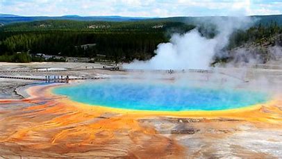
Thermophilic polymerase: Enzyme from heat-loving bacteria (e.g., Taq polymerase).
Sanger DNA Sequencing
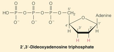
Sanger sequencing uses dideoxynucleotides (ddNTPs) to terminate DNA synthesis at specific bases, allowing determination of DNA sequence.
ddNTPs: Lack 3'-OH, preventing chain extension.
Gel electrophoresis: Separates fragments by size.
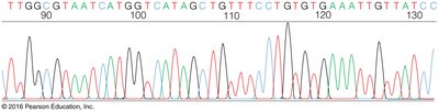
Automated sequencing: Uses fluorescently labeled ddNTPs.
Transcription
Initiation and Elongation
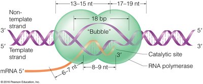
Transcription is the synthesis of RNA from a DNA template. RNA polymerase binds to promoter regions and synthesizes RNA in a transcription bubble.
Promoter: DNA sequence where RNA polymerase binds.
Transcription bubble: Region where DNA is unwound and RNA is synthesized.
σ-factor: Initiates transcription in prokaryotes.
Operon Model and mRNA Fate
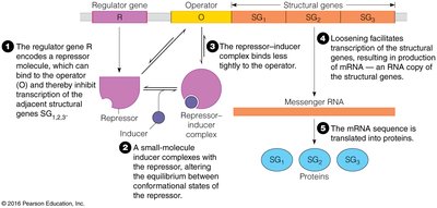
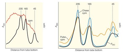
The operon model explains gene regulation in prokaryotes. Messenger RNA (mRNA) is often short-lived and heterogeneous in size.
Regulator gene: Encodes repressor protein.
Operator: DNA region where repressor binds.
Structural genes: Encode proteins.
Inducer: Molecule that inactivates repressor.
Translation
Key Elements of Translation
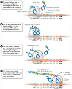
Translation is the process by which ribosomes synthesize proteins using mRNA as a template. The process involves mRNA, tRNA, ribosomes, and various supporting factors.
mRNA: Carries genetic code.
tRNA: Transfers amino acids to ribosome.
Ribosome: Site of protein synthesis.
Aminoacyl-tRNA synthetase: Loads tRNA with amino acids.
Initiation/Elongation factors: Facilitate translation steps.
Molecular chaperones: Assist protein folding.
Codon-Anticodon Pairing and Genetic Code
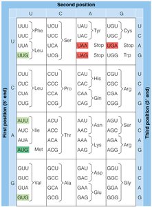
The genetic code is a set of rules by which nucleotide triplets (codons) in mRNA specify amino acids. Codon-anticodon pairing ensures correct translation.
Triplet code: Three nucleotides per codon.
Non-overlapping: Codons are read as distinct units.
No punctuation: Codons are read sequentially without gaps.
Degeneracy: Multiple codons can specify the same amino acid.
Start codon: AUG (methionine or N-formylmethionine).
Stop codons: UAA, UGA, UAG (do not code for amino acids).
tRNA Structure and Function
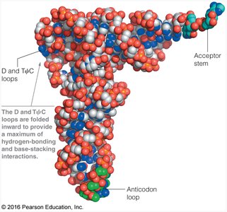
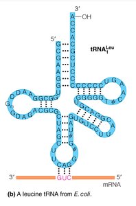
tRNA molecules have a characteristic cloverleaf structure and carry specific amino acids to the ribosome. The anticodon loop pairs with the mRNA codon.
Acceptor stem: Site of amino acid attachment.
Anticodon loop: Pairs with mRNA codon.
D and TψC loops: Provide structural stability.
Ribosome Structure
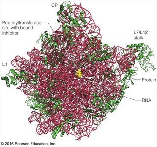
The ribosome is a large ribonucleoprotein complex responsible for protein synthesis. It consists of two subunits and contains rRNA and protein.
Bacterial ribosome: 70S (50S and 30S subunits).
Eukaryotic ribosome: 80S (60S and 40S subunits).
Peptidyl transferase site: Catalyzes peptide bond formation.
Ribozyme: Ribosome's catalytic activity is due to rRNA.
Open Reading Frames and Shine-Dalgarno Sequence
Open reading frames (ORFs) are mRNA regions that can be translated. The Shine-Dalgarno sequence helps align mRNA on the ribosome for translation initiation in prokaryotes.
Start and stop codons: Define the boundaries of ORFs.
Shine-Dalgarno sequence: Located upstream of start codon; complementary to 16S rRNA.
Example: The lac operon mRNA contains multiple ORFs, each with its own Shine-Dalgarno sequence.
*Additional info: The Shine-Dalgarno sequence is essential for proper translation initiation in bacteria, ensuring the ribosome binds at the correct site.*
