 Back
BackGene Expression and Regulation: Foundations of Molecular Genetics
Study Guide - Smart Notes
 Tailored notes based on your materials, expanded with key definitions, examples, and context.
History of DNA Research
Discovery and Identification of Genetic Material
The identification of DNA as the genetic material was a pivotal moment in molecular biology. Early experiments and discoveries laid the foundation for our understanding of heredity and gene expression.
Gregor Mendel (1866): Discovered the basic laws of inheritance using pea plants, establishing the concept of heritable factors (genes).
Friedrich Miescher (1869): Isolated a substance from white blood cells, termed "nuclein," now known as DNA.
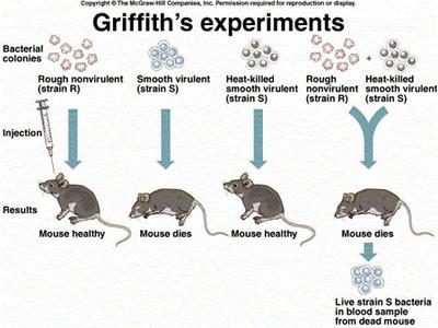
Frederick Griffith (1928): Demonstrated the phenomenon of transformation in bacteria, suggesting the existence of a "transforming principle" that could transfer genetic information.
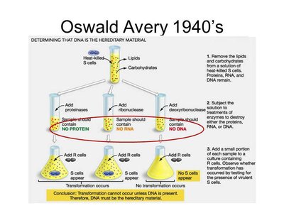
Oswald Avery, Colin MacLeod, and Maclyn McCarty (1944): Identified DNA as the "transforming principle" responsible for heredity, providing strong evidence that DNA is the genetic material.
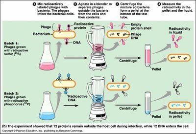
Alfred Hershey and Martha Chase (1952): Used bacteriophages and radioactive labeling to confirm that DNA, not protein, is the genetic material transmitted by viruses to bacteria.
Erwin Chargaff (1947): Established Chargaff’s rules, showing that the amount of adenine equals thymine and cytosine equals guanine in DNA, and that base composition varies between species.
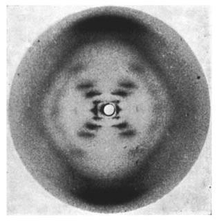
Rosalind Franklin (1950s): Used X-ray crystallography to reveal the helical structure of DNA, providing critical evidence for the double helix model.
James Watson and Francis Crick (1953): Built the first accurate model of DNA’s double helix structure, integrating data from Chargaff and Franklin.
DNA Structure
Nucleotide Composition and Double Helix
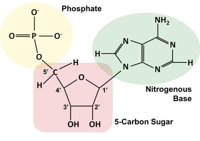
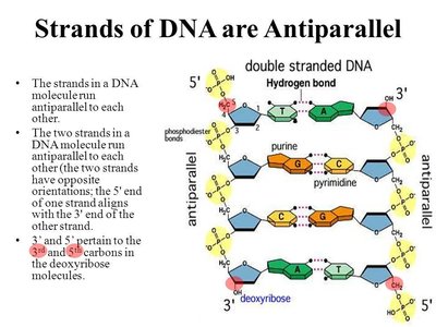
DNA is a polymer composed of nucleotide monomers. Each nucleotide consists of a deoxyribose sugar, a phosphate group, and a nitrogenous base (adenine, thymine, cytosine, or guanine). The structure of DNA is a double helix with two antiparallel strands held together by hydrogen bonds between complementary bases.
Nucleotide Structure: Each nucleotide contains a 5-carbon sugar (deoxyribose), a phosphate group, and a nitrogenous base.
Double Helix: Two strands of nucleotides run in opposite directions (antiparallel), forming a right-handed helix. The backbone is composed of alternating sugars and phosphates, while the bases pair in the center.
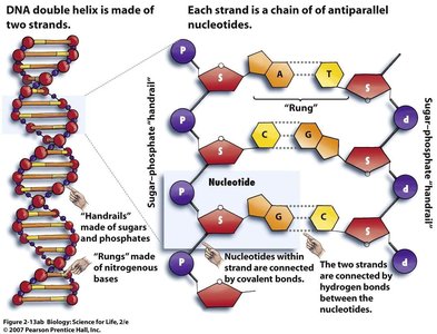
Base Pairing: Adenine pairs with thymine via two hydrogen bonds; cytosine pairs with guanine via three hydrogen bonds. This specificity is crucial for replication and transcription.
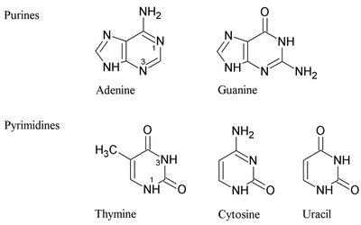
Pyrimidines and Purines: Pyrimidines (cytosine, thymine, uracil) are single-ring structures; purines (adenine, guanine) are double-ring structures. Each base pair consists of one purine and one pyrimidine.
Antiparallel Orientation: One strand runs 5’ to 3’, the other 3’ to 5’. This orientation is essential for DNA replication and function.
DNA Replication
Mechanism and Enzymes
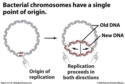
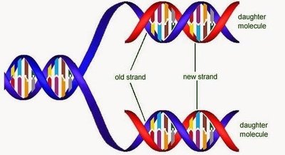
DNA replication is the process by which a cell copies its entire genome before cell division. It is semiconservative, meaning each new DNA molecule consists of one old and one new strand. Replication occurs during the S phase of the cell cycle and involves multiple enzymes and steps.
Origins of Replication: Specific sequences where replication begins. Prokaryotes have a single origin; eukaryotes have multiple origins per chromosome.
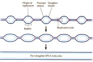
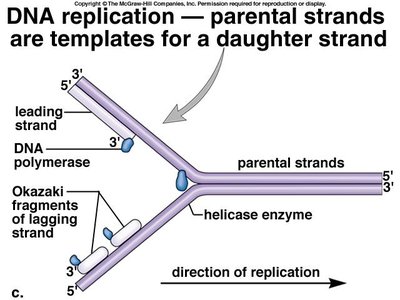
Initiation: Helicase unwinds the DNA, creating replication forks. Single-stranded binding proteins stabilize the unwound DNA. Primase synthesizes short RNA primers to provide a starting point for DNA polymerase.
Elongation: DNA polymerase adds nucleotides in the 5’ to 3’ direction. The leading strand is synthesized continuously, while the lagging strand is synthesized discontinuously in Okazaki fragments, which are later joined by DNA ligase.
Semiconservative Replication: Each daughter DNA molecule contains one parental and one newly synthesized strand.
Proofreading and Repair: DNA polymerase proofreads new DNA, correcting errors. Additional repair mechanisms fix mismatches and other errors.
Telomeres and Telomerase
Chromosome End Protection and Cellular Aging
Telomeres are repetitive, non-coding DNA sequences at the ends of linear chromosomes. They protect coding DNA from degradation during replication. With each cell division, telomeres shorten, eventually leading to cellular senescence or apoptosis. The enzyme telomerase can extend telomeres, active mainly during development and in certain cancer cells.
Telomere Sequence: In humans, the repeat is TTAGGG.
Role in Aging: Telomere shortening limits the number of cell divisions, contributing to aging and acting as a tumor suppressor mechanism.
Telomerase: An enzyme that adds telomeric repeats to chromosome ends, maintaining length in germ cells, stem cells, and cancer cells.
Central Dogma of Molecular Biology
Flow of Genetic Information
The central dogma describes the flow of genetic information from DNA to RNA to protein. This process involves transcription (DNA to RNA) and translation (RNA to protein). Replication ensures genetic continuity. Exceptions include retroviruses (RNA to DNA) and prions (protein to protein).
Replication: DNA is copied to produce identical DNA molecules.
Transcription: DNA is transcribed into messenger RNA (mRNA).
Translation: mRNA is translated into a polypeptide (protein).
Transcription
Process and Regulation
Transcription is the synthesis of RNA from a DNA template. It occurs in three main stages: initiation, elongation, and termination.
Initiation: Transcription factors and RNA polymerase bind to the promoter region (often containing a TATA box) to begin RNA synthesis.
Elongation: RNA polymerase synthesizes a complementary RNA strand in the 5’ to 3’ direction, using one DNA strand as a template.
Termination: RNA polymerase stops transcription at a terminator sequence, releasing the RNA transcript.
RNA Processing and Alternative Splicing
Modification of Eukaryotic mRNA
In eukaryotes, the primary RNA transcript (pre-mRNA) undergoes several modifications before becoming mature mRNA:
5’ Capping: Addition of a protective GTP cap to the 5’ end.
3’ Polyadenylation: Addition of a poly-A tail to the 3’ end for stability and export.
Splicing: Removal of non-coding introns and joining of coding exons by spliceosomes.
Alternative Splicing: Allows different combinations of exons to be joined, producing multiple protein variants from a single gene.
Types of RNA
Functional Diversity
mRNA (Messenger RNA): Encodes amino acid sequences for translation.
tRNA (Transfer RNA): Brings specific amino acids to the ribosome during translation.
rRNA (Ribosomal RNA): Structural and catalytic component of ribosomes.
snRNA (Small nuclear RNA): Involved in RNA splicing in eukaryotes.
Translation
Protein Synthesis
Translation is the process by which ribosomes synthesize proteins using the sequence encoded in mRNA. It occurs in three stages: initiation, elongation, and termination.
Initiation: The small ribosomal subunit binds to mRNA, and the initiator tRNA pairs with the start codon (AUG). The large subunit then assembles.
Elongation: tRNAs bring amino acids to the ribosome, matching their anticodons to mRNA codons. Peptide bonds form between amino acids, elongating the polypeptide chain.
Termination: When a stop codon is reached, release factors promote the release of the completed polypeptide and disassembly of the ribosome.
Wobble Effect: The third base of a codon can often vary without changing the amino acid specified, allowing fewer tRNAs to recognize multiple codons and providing protection against mutations.
Post-Translational Modification and Protein Folding
Functional Maturation of Proteins
Folding: Newly synthesized polypeptides fold into their functional three-dimensional structures, often with the help of chaperone proteins.
Modifications: Proteins may undergo glycosylation, lipidation, phosphorylation, or ubiquitination, affecting their function, localization, or stability.
Mutations and Genetic Disorders
Types and Consequences of Mutations
Point Mutations: Single nucleotide changes, including silent (no effect), missense (amino acid change), and nonsense (premature stop codon) mutations.
Frameshift Mutations: Insertions or deletions that alter the reading frame, often resulting in nonfunctional proteins.
Genetic Disorders: Examples include sickle cell anemia (missense mutation) and Tay Sachs disease (frameshift mutation).
Somatic vs. Germline Mutations: Germline mutations are heritable; somatic mutations affect only the individual.
Gene Regulation
Control of Gene Expression
Gene regulation ensures that genes are expressed only when needed, allowing cellular differentiation and adaptation.
Operons (Prokaryotes): Groups of genes regulated together. Inducible operons (e.g., lac operon) are usually off but can be turned on; repressible operons (e.g., trp operon) are usually on but can be turned off.
Eukaryotic Regulation: Occurs at multiple levels, including chromatin structure (euchromatin vs. heterochromatin), transcriptional control (transcription factors), post-transcriptional control (RNA processing), translational control (microRNAs), and post-translational control (protein modification and degradation).
Epigenetics: Heritable changes in gene expression not involving changes in DNA sequence, such as DNA methylation and histone acetylation.
Biotechnology and Genetic Engineering
Techniques and Applications
DNA Cloning: Inserting DNA fragments into plasmids and propagating them in bacteria to produce multiple copies or express proteins.
Polymerase Chain Reaction (PCR): Amplifies specific DNA sequences using cycles of denaturation, annealing, and extension.
Gel Electrophoresis: Separates DNA, RNA, or proteins based on size and charge for analysis.
DNA Sequencing: Determines the order of nucleotides in DNA, enabling gene identification and comparative genomics.
CRISPR/Cas9: A genome editing tool that allows precise modifications of DNA sequences in living organisms.
Summary Table: Key Differences Between DNA and RNA
Feature | DNA | RNA |
|---|---|---|
Sugar | Deoxyribose | Ribose |
Bases | A, T, C, G | A, U, C, G |
Strands | Double-stranded | Single-stranded |
Location | Nucleus (eukaryotes) | Nucleus & cytoplasm |
Function | Genetic information storage | Protein synthesis, regulation |
Summary Table: Types of Mutations
Mutation Type | Description | Effect |
|---|---|---|
Silent | Base change, no amino acid change | No effect |
Missense | Base change, different amino acid | Variable (e.g., sickle cell anemia) |
Nonsense | Base change, premature stop codon | Truncated, nonfunctional protein |
Frameshift | Insertion/deletion not in multiples of 3 | Severe, often nonfunctional protein |
