 Back
BackImmunological Techniques in Biochemistry: Antibody-Based Methods and Applications
Study Guide - Smart Notes
 Tailored notes based on your materials, expanded with key definitions, examples, and context.
Immunological Techniques in Biochemistry
Antibodies and Antigens
Antibodies are specialized proteins produced by the immune system to recognize and bind to foreign molecules called antigens. This specificity makes antibodies invaluable tools in biochemistry research for detecting, quantifying, and isolating biomolecules.
Antibodies (Immunoglobulins): Proteins with high specificity for antigens; produced in animals for research purposes.
Antigen: Any foreign molecule recognized by antibodies; most antigens are proteins, but can also include carbohydrates, nucleic acids, and lipids.
Epitope: The specific region of an antigen recognized by an antibody. Epitopes can be linear (continuous amino acid sequence) or conformational (formed by protein folding).
Antibody Classes: IgG, IgM, IgA, IgE, IgD; IgG is most abundant and widely used in research.
Example: Mouse anti-actin antibody binds specifically to actin protein; actin is the antigen, and the recognized sequence is the epitope.
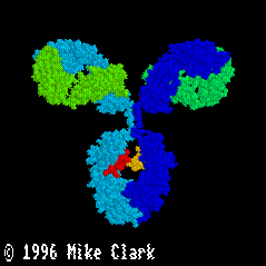
Antibody Structure
Antibodies are tetrameric glycoproteins composed of two identical heavy chains and two identical light chains, connected by disulfide bonds. The structure is often depicted as a "Y" shape.
Domains: Variable (VL, VH) and constant (CL, CH1, CH2, CH3) domains; variable domains are responsible for antigen binding.
Antigen Binding Regions: Each antibody has two identical antigen binding sites formed by VL and VH domains.
Glycoprotein Nature: Heavy chains are bound to oligosaccharides.
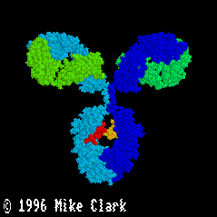
Preparation of Antibodies for Research
Antibodies are generated by immunizing animals with purified antigens or synthetic peptides. The resulting serum contains polyclonal antibodies, which recognize multiple epitopes. Monoclonal antibodies, produced by a single B cell clone, recognize a single epitope.
Immunization: Antigen injected into animals (mice, rabbits, etc.) via various routes (i.p., i.d., i.v., i.m., s.c.).
Booster Immunizations: Increase antibody affinity and concentration.
Antiserum: Serum containing antibodies, used directly or purified.
Polyclonal vs. Monoclonal: Polyclonal antibodies recognize multiple epitopes; monoclonal recognize one.
Primary and Secondary Antibodies
Primary antibodies bind directly to the antigen. Secondary antibodies, produced in a different species, bind to primary antibodies and are often conjugated to detectable labels (enzymes or fluorophores).
Primary Antibody: Binds to the target antigen.
Secondary Antibody: Binds to primary antibody; used for detection and amplification.
Labeling: Secondary antibodies are labeled with enzymes (e.g., HRP) or fluorophores (e.g., FITC, TRITC).
Antibody Labels
Labels attached to secondary antibodies enable detection via colorimetric, chemiluminescent, or fluorescent methods.
Enzyme Labels: HRP and alkaline phosphatase produce colored or luminescent products.
Fluorophores: FITC (green), TRITC (red/orange), DAPI (blue, binds DNA).
Summary of Immunological Techniques
Antibody-based techniques are used to detect, quantify, and localize proteins, diagnose diseases, count and isolate cells, and study protein interactions.
Indirect ELISA: Detects antibodies to a specific antigen.
Sandwich ELISA: Detects a specific antigen in samples.
Western Blotting: Detects and quantifies proteins in samples.
Immunofluorescence Microscopy: Localizes proteins in cells.
Flow Cytometry and FACS: Counts and isolates specific cell types.
Immunoaffinity Chromatography: Purifies proteins from mixtures.
Immunoprecipitation: Identifies protein-protein interactions.
Antibody-Based Techniques
Enzyme-Linked Immunosorbent Assay (ELISA)
ELISA is a plate-based assay used for detecting antibodies (indirect ELISA) or antigens (sandwich ELISA) in biological samples. It is widely used in medical diagnostics and research.
Indirect ELISA: Detects antibodies to a specific antigen (e.g., HIV gp120) in blood samples.
Sandwich ELISA: Detects antigens (e.g., malaria protein HrpII) using two antibodies recognizing different epitopes.
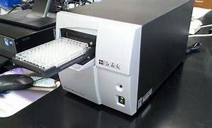
Detection: Color development measured by absorbance in a plate reader.
Western Blotting
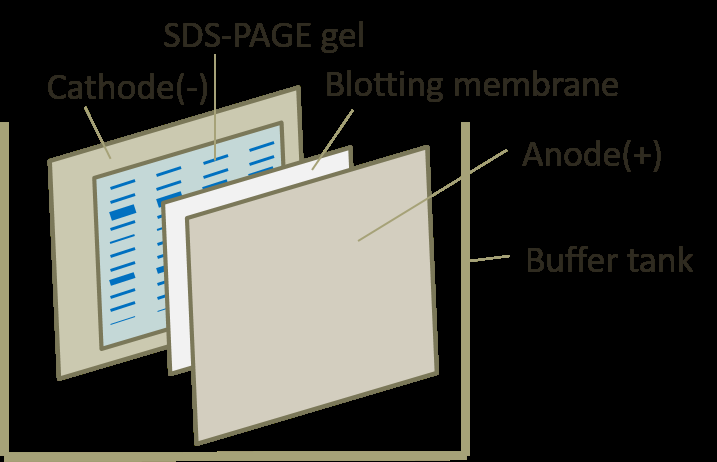
Western blotting is used to detect and quantify specific proteins in samples. Proteins are separated by SDS-PAGE, transferred to a membrane, and detected using primary and secondary antibodies.
SDS-PAGE: Separates proteins by molecular weight.
Transblotting: Transfers proteins from gel to membrane using an electric field.
Detection: Chemiluminescent substrate produces light, photographed for analysis.

Immunofluorescence Microscopy
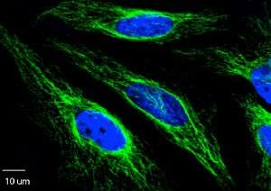
This technique localizes specific proteins within cells using fluorophore-labeled antibodies and fluorescence microscopy. It is useful for studying protein distribution and co-localization in cellular compartments.
Sample Preparation: Cells are fixed and permeabilized to allow antibody access.
Primary Antibody: Binds to target protein.
Secondary Antibody: Conjugated to fluorophore, binds to primary antibody.
Microscopy: Fluorescence microscope visualizes labeled proteins.
Coverslip Handling and Cell Culture
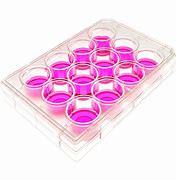
Coverslips are used to grow cells for immunofluorescence experiments. After staining, coverslips are mounted on microscope slides for imaging.
Coverslip Preparation: Cells are cultured on coverslips, fixed, and stained.
Mounting: Coverslips are placed face-down on slides for microscopy.


Co-localization in Immunofluorescence Microscopy
Co-localization allows simultaneous visualization of two or more proteins in the same cell using antibodies from different species and fluorophores with distinct emission wavelengths.
Multiple Antibodies: Use primary antibodies from different animals and secondary antibodies conjugated to different fluorophores.
Imaging: Separate images are merged to show co-localization.
Organelle Marker Proteins: Used to confirm protein localization in specific organelles.
Flow Cytometry and FACS
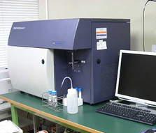
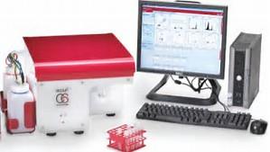
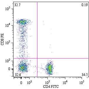
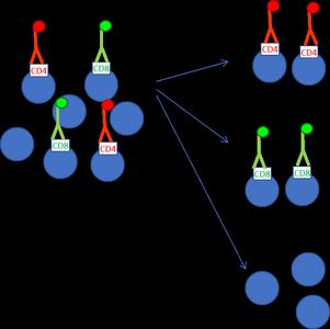
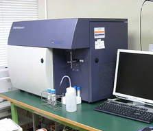
Flow cytometry counts and analyzes cells labeled with fluorophore-conjugated antibodies. FACS (Fluorescence Activated Cell Sorting) isolates specific cell types based on fluorescence.
Cell Marker Proteins: Used to identify cell types (e.g., CD4 for Th cells, CD8 for Tc cells).
Counting Cells: Flow cytometer measures fluorescence intensity to count cell populations.
FACS: Sorts cells into separate tubes based on fluorescence.
Immunoaffinity Chromatography
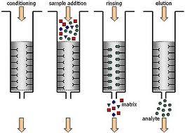
Immunoaffinity chromatography is used to purify specific proteins from complex mixtures using antibody-coated beads in a column. The target protein binds to the antibody, is washed, and then eluted under acidic conditions.
Antibody-Coated Beads: Agarose beads cross-linked to antibodies or protein A.
Column Preparation: Protein sample passed through column; target protein binds to beads.
Elution: Bound protein released by acidic buffer.
Immunoprecipitation
Immunoprecipitation isolates a protein and its binding partners from cell lysates using antibody-coated beads. The proteins are released by SDS-PAGE buffer and analyzed by gel electrophoresis and mass spectrometry.
Procedure: Antibody-coated beads mixed with cell lysate; target protein and interacting proteins bind to beads.
Pelleting: Beads are pelleted by centrifugation, washed, and proteins released by SDS-PAGE buffer.
Analysis: SDS-PAGE separates proteins; mass spectrometry identifies binding partners.
Conclusion
Immunological techniques are essential tools in biochemistry for studying protein structure, function, localization, and interactions. Mastery of these methods enables researchers to diagnose diseases, analyze cellular processes, and purify biomolecules for further study.
