 Back
Lipids and Biological Membranes: Structure, Function, and Classification
Study Guide - Smart Notes
 Tailored notes based on your materials, expanded with key definitions, examples, and context.
Lipids: Classification and Properties
Overview of Lipid Classes
Lipids are a diverse group of biological molecules characterized by their solubility in nonpolar solvents and poor solubility in water. They play essential roles in energy storage, membrane structure, and signaling.
Fatty Acids: Long-chain hydrocarbons with a terminal carboxylic acid group.
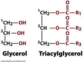
Triacylglycerols: Esters of glycerol with three fatty acids, major energy storage molecules.
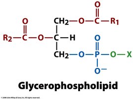
Glycerophospholipids: Major components of biological membranes, containing a glycerol backbone, two fatty acids, and a phosphorylated head group.
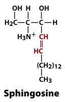
Sphingolipids: Membrane lipids with a sphingosine backbone.
Isoprenoids: Lipids derived from isoprene units, including cholesterol and vitamins.
Cholesterol: A steroid important for membrane structure and precursor to steroid hormones.
Fatty Acids: Structure and Types
Fatty acids are amphiphilic molecules with a polar carboxyl head and a nonpolar hydrocarbon tail. They can be classified as saturated, unsaturated, or polyunsaturated based on the presence and number of double bonds.
Saturated fatty acids: No double bonds; straight chains allow tight packing and higher melting points.
Unsaturated fatty acids: One or more double bonds; kinks in the chain reduce packing and lower melting points.
Polyunsaturated fatty acids: Multiple double bonds; important for membrane fluidity and signaling.
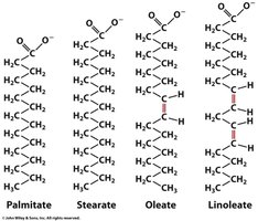
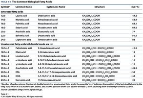
Table: Common Biological Fatty Acids
Symbol | Systematic Name | Structure | mp (°C) |
|---|---|---|---|
12:0 | Lauric acid | CH3(CH2)10COOH | 44.2 |
16:0 | Palmitic acid | CH3(CH2)14COOH | 63.1 |
18:0 | Stearic acid | CH3(CH2)16COOH | 69.6 |
18:1 | Oleic acid | CH3(CH2)7CH=CH(CH2)7COOH | 13.4 |
18:2 | Linoleic acid | CH3(CH2)4CH=CHCH2CH=CH(CH2)6COOH | -5 |
20:4 | Arachidonic acid | CH3(CH2)4(CH=CHCH2)4(CH2)2COOH | -49 |
Triacylglycerols: Structure and Function
Triacylglycerol Structure
Triacylglycerols (triglycerides) are formed by esterification of three fatty acids to a glycerol backbone. They serve as the primary energy storage molecules in animals.
Energy Density: Fats provide about six times the metabolic energy of an equal weight of hydrated glycogen.
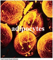
Adipocytes: Specialized cells for synthesis and storage of triacylglycerols.
Glycerophospholipids: Membrane Structure
Structure and Amphiphilicity
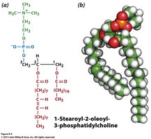
Glycerophospholipids are major lipid components of biological membranes. They consist of a glycerol backbone, two fatty acid tails, and a phosphorylated head group (X). Their amphiphilic nature allows them to form bilayers.
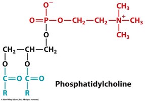
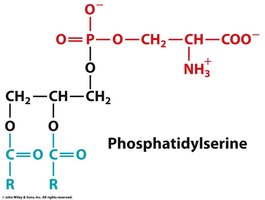
Head Groups: Common head groups include choline, serine, and ethanolamine.
Function: Structural foundation of cell membranes.
Common Glycerophospholipid Head Groups
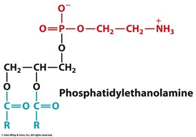
Phosphatidylcholine: Most abundant in eukaryotic membranes.
Phosphatidylserine: Important for cell signaling and apoptosis.
Phosphatidylethanolamine: Contributes to membrane curvature and flexibility.
Phospholipases: Enzymatic Cleavage
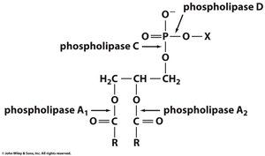
Phospholipases are enzymes that cleave phospholipids at specific sites, playing roles in membrane remodeling and signal transduction.
Phospholipase A1 and A2: Remove fatty acids from the glycerol backbone.
Phospholipase C: Cleaves before the phosphate group.
Phospholipase D: Cleaves after the phosphate group.
Plasmalogens: Ether-Linked Glycerophospholipids
Plasmalogens are a subclass of glycerophospholipids where the C1 substituent is linked via an unsaturated ether bond instead of an ester bond. They are abundant in heart and brain tissues. 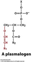
Sphingolipids: Structure and Function
Sphingosine Backbone
Sphingolipids are major membrane components with significant physiological roles. Unlike glycerophospholipids, they use sphingosine as a backbone. 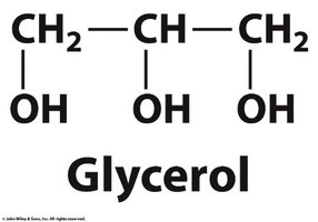
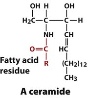
Ceramides and Sphingomyelins
Ceramide: Formed by attaching a fatty acid to sphingosine.
Sphingomyelin: Most common sphingolipid, contains phosphocholine or phosphoethanolamine head group; found in myelin sheath of nerves.
Glycosphingolipids: Cerebrosides and Gangliosides
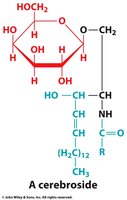
Cerebrosides: Sphingolipids with monosaccharide head groups; important in brain and nerve tissue.
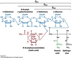
Gangliosides: Sphingolipids with oligosaccharide head groups; act as cell-surface receptors and determinants of cell-cell recognition.
Cholesterol and Steroid Lipids
Cholesterol: Structure and Function
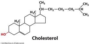
Cholesterol is the most abundant steroid in animals and a major component of plasma membranes. Its fused ring structure provides rigidity and it is a precursor for steroid hormones.
Amphiphilic: Contains both polar and nonpolar regions.
Hormone precursor: Metabolic precursor for cortisol, aldosterone, testosterone, and estrogen.
Vitamin D: Calcium Metabolism
Vitamin D is a sterol derivative hormone regulating calcium metabolism. Its active forms are produced by enzymatic hydroxylation in the liver and kidney.
Function: Increases intestinal absorption of Ca2+, promoting bone and teeth deposition.
Other Isoprenoid Lipids
Ubiquinone (Coenzyme Q): Electron carrier in mitochondrial membrane.
Vitamin K: Essential for blood clotting; synthesized by plants and bacteria.
Vitamin A (Retinol): Derived from β-carotene; functions as a photoreceptor in the eye.
Vitamin E: Antioxidant protecting membrane lipids and proteins.
Eicosanoids: Signaling Lipids
Origin and Function
Eicosanoids are derived from arachidonic acid and act as local signaling molecules.
Roles: Regulation of pain, fever, blood pressure, coagulation, and reproduction.
Lipid Aggregates and Membrane Structure
Micelles, Liposomes, and Bilayers
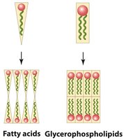
Amphiphilic lipids aggregate in aqueous solutions to form micelles, liposomes, or bilayers, depending on their structure.
Micelles: Formed by single-tailed lipids (e.g., fatty acids).
Liposomes: Self-sealing vesicles formed by phospholipids.
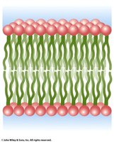
Lipid Bilayers: Structural basis of biological membranes.
Bilayer Characteristics
Fluidity: Lipid bilayers are fluid, with head groups and tails in constant motion.
Asymmetry: Different lipids are found in each leaflet of the bilayer.
Membrane Diffusion
Lipid molecules diffuse within membranes either laterally (rapid) or transversely (flip-flop, very slow). 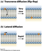
Temperature Dependence
Transition Temperature: Below this, bilayers become gel-like solids.
Chain Length and Saturation: Longer and more saturated chains increase melting point and decrease fluidity.
Membrane Proteins: Types and Functions
Association with Membranes
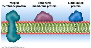
Proteins associate with membranes as integral, peripheral, or lipid-linked proteins.
Integral (Intrinsic) Proteins: Tightly associated via hydrophobic effects; span the bilayer.
Peripheral (Extrinsic) Proteins: Loosely associated; can be dissociated by mild treatments.
Lipid-Linked Proteins: Covalently attached to lipids; anchor proteins to membranes.
Transmembrane Protein Structure
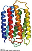
Alpha Helices: Common in integral proteins (e.g., bacteriorhodopsin).
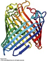
Beta Barrels: Found in porins, channel-forming proteins.
Lipid-Linked Proteins
Myristoylation, Palmitoylation, Prenylation: Types of lipid modifications anchoring proteins to membranes.
Peripheral Proteins
Peripheral proteins do not bind lipids and behave as water-soluble proteins. Example: cytochrome c, associated with the mitochondrial membrane. 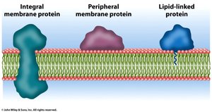
Fluid Mosaic Model and Membrane Asymmetry
Fluid Mosaic Model
The fluid mosaic model describes membranes as a dynamic structure where proteins float in a sea of lipids, allowing lateral diffusion. 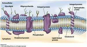
Phospholipid Distribution
Membranes have asymmetric distribution of phospholipids between inner and outer leaflets, contributing to functional specialization. 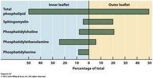
Summary Table: Lipid Classes and Functions
Lipid Class | Main Function | Example |
|---|---|---|
Fatty Acids | Energy storage, membrane structure | Palmitate, Oleate |
Triacylglycerols | Energy reservoir | Triglycerides in adipocytes |
Glycerophospholipids | Membrane structure | Phosphatidylcholine |
Sphingolipids | Membrane structure, signaling | Sphingomyelin, Ganglioside |
Cholesterol | Membrane rigidity, hormone precursor | Cholesterol |
Isoprenoids | Electron transport, vitamins | Ubiquinone, Vitamin K |
Additional info: Academic context was added to clarify the roles and structures of lipid classes, membrane proteins, and the fluid mosaic model. All images included are directly relevant to the adjacent explanations and reinforce key concepts in biochemistry.
