 Back
BackLipids, Membranes, and Cellular Transport: Structure, Function, and Regulation
Study Guide - Smart Notes
 Tailored notes based on your materials, expanded with key definitions, examples, and context.
Lipids: Structure and Function
Overview of Lipids
Lipids are a diverse group of water-insoluble organic molecules that play critical roles in biological systems. Their functions include forming structural components of membranes, serving as energy storage molecules, acting as vitamins, and mediating chemical signaling and regulation.
Structural diversity: Lipids vary in their hydrocarbon chain length, degree of saturation, and functional groups.
Functional diversity: Lipids are involved in membrane structure, energy storage, insulation, and signaling.
Fatty Acids: Structure and Classification
Fatty acids are carboxylic acids with long aliphatic hydrocarbon tails. They are fundamental building blocks of many complex lipids.
General structure: A polar carboxyl (COOH) head group and a nonpolar hydrocarbon tail.
pKa: 4.5–5.0; at physiological pH (~7), fatty acids are deprotonated (anion form, name ends in 'ate').
Variations: Tail length, number and position of double bonds, and branching.
Saturated vs. Unsaturated Fatty Acids
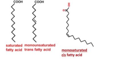
Saturated fatty acids: No C–C double bonds; tails are saturated with hydrogen atoms.
Unsaturated fatty acids: Contain one or more C–C double bonds.
Monounsaturated: One double bond.
Polyunsaturated: Multiple double bonds.
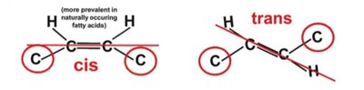
Double bond configuration: Naturally occurring fatty acids are mostly in the cis configuration, which introduces a kink in the hydrocarbon chain, affecting physical properties such as melting temperature.
Physical Properties and Melting Temperature
Tail length: Longer tails increase melting temperature (Tm).
Double bonds: Presence of double bonds (especially cis) lowers Tm, making unsaturated fatty acids liquid at room temperature (oils), while saturated fatty acids are typically solid.
Fatty Acid Nomenclature
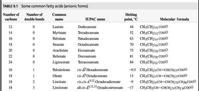
Common names: Do not indicate structure directly (e.g., laurate, palmitate).
IUPAC names: Indicate number of carbons and double bonds (e.g., hexadecanoate for palmitate).
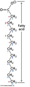
Shorthand notation: Number of carbons:number of double bonds Δ(position), e.g., 18:2 Δ9,12 for linoleate.
Greek letter system: The carbon next to the carboxyl is α, terminal carbon is ω; ω-3 fatty acids have a double bond three carbons from the end.
Fatty Acid Structure Example
Essential Fatty Acids
Essential fatty acids cannot be synthesized by the body and must be obtained from the diet. These include omega-3 (ω-3) and omega-6 (ω-6) fatty acids, which are important for cardiovascular health and cellular function.
Complex Lipids: Triacylglycerols and Glycerophospholipids
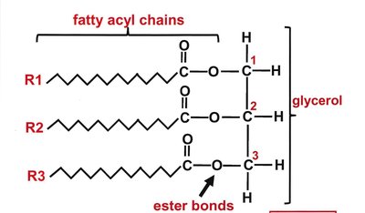
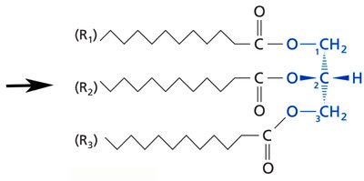
Triacylglycerols (Triglycerides)
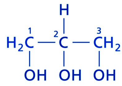
Triacylglycerols are the main storage form of energy in animals. They consist of three fatty acids esterified to a glycerol backbone.
Structure: Three fatty acyl chains attached to glycerol via ester bonds.
Function: Long-term energy storage, thermal insulation, and metabolic energy source.
Physical properties: Hydrophobic; physical state (solid or liquid) depends on fatty acid composition.
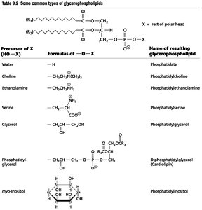
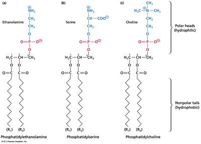
Glycerophospholipids (Phosphoglycerides)
Glycerophospholipids are major components of biological membranes. They contain two fatty acids and a phosphate group attached to a glycerol backbone.
Structure: Glycerol backbone, two fatty acyl chains (R1 usually saturated, R2 unsaturated), and a phosphate group with various head groups.
Amphipathic nature: Hydrophobic tails and hydrophilic head group enable membrane formation.
Membrane Formation
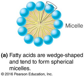
Micelles: Fatty acids form spherical micelles due to their wedge shape.
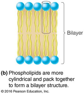
Bilayers: Phospholipids form bilayers, the fundamental structure of cell membranes.
Sterols and Cholesterol
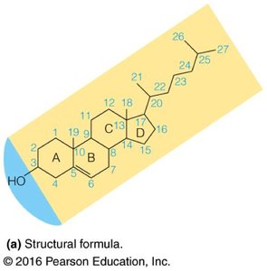
Cholesterol: Structure and Function
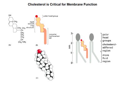
Cholesterol is a rigid, hydrophobic molecule essential for membrane structure and function. It modulates membrane fluidity and serves as a precursor for steroid hormones and bile acids.
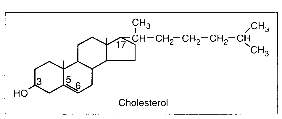
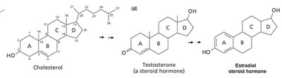
Structure: Four fused rings (A, B, C, D) with a hydrocarbon tail and a hydroxyl group at C3.
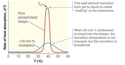
Membrane role: Increases membrane fluidity at low temperatures and decreases it at high temperatures, broadening the phase transition.
Precursor: Steroid hormones (e.g., testosterone, estradiol) and bile salts.
Lipoproteins and Lipid Transport
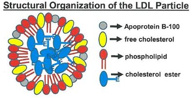
Lipoprotein Particles
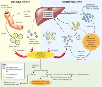
Lipoproteins are complexes of lipids and proteins that transport hydrophobic lipids through the aqueous bloodstream. Major classes include chylomicrons, LDL, and HDL, each with distinct roles in lipid transport and metabolism.
Chylomicrons: Transport dietary triacylglycerols from intestines to tissues.
Low-density lipoprotein (LDL): Delivers cholesterol to peripheral tissues; high levels are associated with cardiovascular risk.
High-density lipoprotein (HDL): Collects cholesterol from tissues and returns it to the liver for excretion; high levels are protective.
Biological Membranes and Membrane Proteins
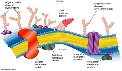
Membrane Structure and Protein Association
Biological membranes are composed of lipid bilayers with embedded proteins. Proteins associate with membranes in three main ways:
Integral (transmembrane) proteins: Span the bilayer, interacting with both hydrophobic core and aqueous environments.
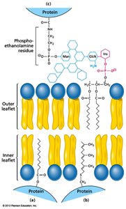
Lipid-anchored proteins: Covalently attached to lipid molecules that insert into the membrane.
Peripheral proteins: Associate with membrane surfaces via non-covalent interactions with lipid head groups or other proteins.
Membrane Transport Mechanisms
Types of Membrane Transport
Membranes regulate the movement of substances into and out of cells via several mechanisms:
Simple diffusion: Passive movement down a concentration gradient without protein assistance.
Facilitated diffusion: Passive, protein-mediated transport (e.g., channels, carriers).
Active transport: Protein-mediated movement against a gradient, requiring energy (ATP, ion gradients, or light).

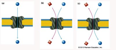
Transporter Classification
Uniport: Transports one substrate at a time.
Symport: Transports two substrates in the same direction.
Antiport: Transports two substrates in opposite directions.
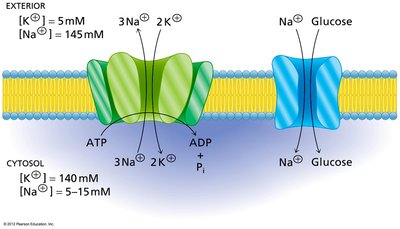
Active Transport Example
The Na+/K+ ATPase is a primary active transporter that uses ATP to pump Na+ out and K+ into the cell, establishing electrochemical gradients essential for cellular function. Secondary active transporters use these gradients to drive the uptake of other molecules, such as glucose.
Signal Transduction and Membrane Dynamics
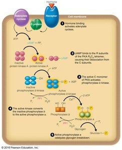
General Model of Signal Transduction
Cells respond to external stimuli via signal transduction pathways, which typically involve membrane receptors, transducers, effectors, and second messengers. These cascades regulate diverse cellular processes, including metabolism, growth, and gene expression.
First messenger: Extracellular signal (e.g., hormone).
Receptor: Membrane protein that binds the signal.
Transducer: Protein that relays the signal (e.g., G protein).
Effector: Enzyme that generates a second messenger (e.g., adenylyl cyclase).
Second messenger: Small molecule that propagates the signal inside the cell (e.g., cAMP, Ca2+).
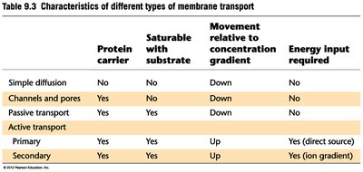
Summary Table: Types of Membrane Transport
Type | Protein Carrier | Saturable with Substrate | Movement Relative to Gradient | Energy Input Required |
|---|---|---|---|---|
Simple diffusion | No | No | Down | No |
Channels and pores | Yes | No | Down | No |
Passive transport | Yes | Yes | Down | No |
Active transport (primary) | Yes | Yes | Up | Yes (direct source) |
Active transport (secondary) | Yes | Yes | Up | Yes (on gradient) |
Additional info: This guide covers the structure, classification, and function of lipids, their role in membrane structure, the mechanisms of membrane transport, and the basics of signal transduction, as relevant to a college-level biochemistry course.
