 Back
BackMembranes, Membrane Proteins, and Transport Mechanisms
Study Guide - Smart Notes
 Tailored notes based on your materials, expanded with key definitions, examples, and context.
Membrane Structure and Composition
Membrane Lipid Diversity
Biological membranes are composed of a diverse array of lipids, whose composition varies between organisms, tissues, and organelles. The ratio of lipid to protein, the types of phospholipids, and the abundance of sterols are all variable features that contribute to membrane function and specialization.
Phospholipids: Major structural components of membranes, including glycerophospholipids and sphingolipids.
Sterols: Cholesterol is predominant in animal plasma membranes, while other sterols (e.g., ergosterol, stigmasterol) are found in yeast and plants.
Glycolipids: Abundant in plant chloroplasts and certain animal tissues.
Membrane Asymmetry: Lipid and protein distribution differs between the inner and outer leaflets of the bilayer.
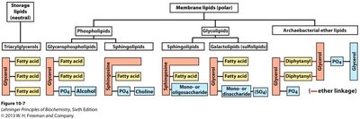
Major Membrane Lipid Classes
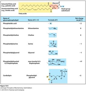
Glycerophospholipids: Built on a glycerol backbone, with two fatty acids and a phosphate-containing head group.
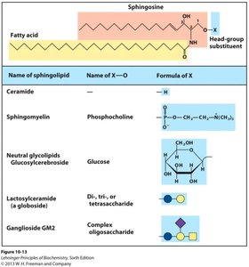
Sphingolipids: Built on a sphingosine backbone, with a fatty acid and a polar head group (e.g., phosphocholine or sugars).
Glycolipids: Lipids with carbohydrate groups, important for cell recognition.
Archaeal Ether Lipids: Unique to archaea, with ether linkages and branched chains.
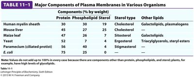
Membrane Composition in Different Organisms
The composition of plasma membranes varies significantly among different organisms and cell types, reflecting adaptation to specific functional requirements.
Organism | Protein (%) | Phospholipid (%) | Sterol (%) | Sterol Type | Other Lipids |
|---|---|---|---|---|---|
Human myelin sheath | 30 | 30 | 19 | Cholesterol | Galactolipids, plasmalogens |
Mouse liver | 45 | 27 | 25 | Cholesterol | — |
Maize leaf | 47 | 26 | 7 | Sitosterol | Galactolipids |
Yeast | 52 | 7 | 7 | Ergosterol | Triacylglycerols, steryl esters |
Paramecium | 56 | 40 | 2 | Stigmasterol | — |
E. coli | 75 | 25 | 0 | — | — |
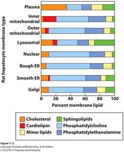
Distribution of Lipids in Cellular Membranes
Different cellular membranes have distinct lipid compositions, which are crucial for their specific functions. For example, the plasma membrane is rich in cholesterol and sphingolipids, while the inner mitochondrial membrane contains cardiolipin.
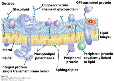
Membrane Structure: The Fluid Mosaic Model
Organization of Membrane Components
The fluid mosaic model describes the membrane as a dynamic, two-dimensional liquid where lipids and proteins can move laterally. Membrane proteins are embedded within or associated with the lipid bilayer, contributing to a variety of cellular functions.
Integral proteins: Span the membrane, often as alpha-helices or beta-barrels.
Peripheral proteins: Loosely associated with the membrane surface.
Lipid-anchored proteins: Covalently attached to membrane lipids.
Carbohydrates: Present only on the extracellular side, attached to lipids (glycolipids) or proteins (glycoproteins).

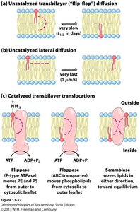
Membrane Lipid Mobility and Asymmetry
Lipids can move laterally within the leaflet rapidly, but transbilayer (flip-flop) movement is slow and often requires enzymes such as flippases, floppases, and scramblases. Membrane asymmetry is maintained by these enzymes and is essential for function.
Lipid Rafts
Lipid rafts are microdomains within the membrane, enriched in cholesterol, sphingolipids, and certain proteins. They are more ordered and less fluid than the surrounding membrane, serving as platforms for signaling and protein sorting.
Membrane Curvature and Fusion
Mechanisms of Membrane Curvature
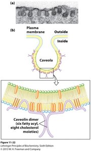
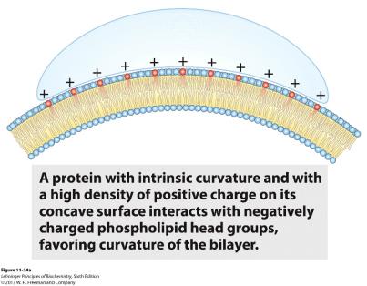
Membrane curvature is essential for processes such as vesicle formation, fusion, and fission. Proteins can induce curvature by various mechanisms, including insertion of amphipathic helices, scaffolding, or clustering of lipids.
Caveolin: An integral membrane protein that induces curvature by forming oligomers and inserting acyl chains into the membrane.
BAR domain proteins: Polymerize to stabilize curved membrane regions.wtf is even this
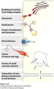
Membrane Fusion
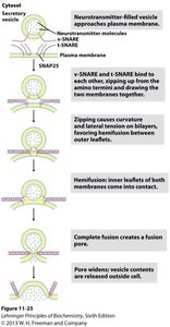
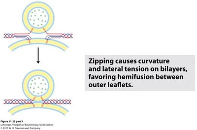
Membrane fusion is a fundamental process in exocytosis, endocytosis, fertilization, and viral entry. It involves the merging of two lipid bilayers into one, mediated by specialized proteins (e.g., SNAREs) that bring membranes into close proximity and induce hemifusion followed by full fusion.
Transport Across Membranes
Types of Membrane Transport
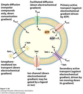
Transport across biological membranes is essential for nutrient uptake, waste removal, and signal transduction. Transport mechanisms are classified based on energy requirements and specificity.
Simple diffusion: Passive movement of small, nonpolar molecules down their concentration gradient.
Facilitated diffusion: Passive transport of molecules via specific carrier proteins or channels.
Active transport: Movement of molecules against their concentration gradient, requiring energy (e.g., ATP hydrolysis).
Energetics of Membrane Transport
The free energy change () for transport depends on the concentration gradient and, for charged molecules, the membrane potential. The Nernst equation describes the equilibrium potential for an ion across a membrane.
Fick's Law: , where is the rate of diffusion, is the permeability coefficient, and is the concentration difference.
Electrochemical potential:
Nernst Equation:
Carrier Proteins and Channels
Transport proteins facilitate the movement of specific molecules across membranes. Carriers (permeases, transporters) undergo conformational changes to move solutes, while channels form aqueous pores for rapid ion movement.
Facilitated diffusion: Specific, saturable, and much faster than simple diffusion. Example: Glucose transporter (GLUT1).
Active transport: Includes P-type ATPases (e.g., Na+/K+ ATPase, Ca2+ ATPase) and light-driven pumps (e.g., bacteriorhodopsin).
Ion Channels and Membrane Potentials
Ion Channel Types and Function
Ion channels are integral membrane proteins that allow selective and rapid movement of ions across membranes. They are essential for generating membrane potentials and signal transduction.
Voltage-gated channels: Open in response to changes in membrane potential.
Ligand-gated channels: Open in response to binding of signaling molecules (e.g., neurotransmitters).
Mechanically-gated channels: Open in response to mechanical force.
Membrane Potential and the Nernst Equation
The membrane potential is determined by the distribution of ions and the selective permeability of the membrane. The Nernst equation predicts the equilibrium potential for a given ion.
Membrane potential (): The voltage difference across the membrane, typically negative inside relative to outside.
Nernst equation (simplified at 20°C for monovalent cations):
Membrane Receptors and Signal Transduction
Receptor Specificity and Ligand Binding
Membrane receptors are proteins that bind specific extracellular signaling molecules (ligands) and initiate cellular responses. The specificity of receptors allows cells to respond selectively to different signals.
Ligands: Include ions, hormones, peptides, and proteins.
Affinity: Measured by the dissociation constant (); lower indicates higher affinity.
G-Protein Coupled Receptors (GPCRs)
GPCRs are a large family of membrane receptors that mediate responses to hormones, neurotransmitters, and sensory stimuli. They activate heterotrimeric G-proteins, which in turn regulate various effectors such as enzymes and ion channels.
Second messengers: Small molecules like cAMP that propagate the signal inside the cell.
Example: Epinephrine receptor mediates the fight-or-flight response by activating glycogen breakdown and increasing heart rate.
Receptor Tyrosine Kinases (RTKs)
RTKs are membrane receptors with intrinsic kinase activity. Ligand binding induces receptor dimerization and autophosphorylation, triggering downstream signaling cascades that regulate cell growth, differentiation, and metabolism.
Example: Insulin receptor activates a kinase cascade leading to increased glucose uptake and gene expression.
Summary Table: Key Membrane Concepts
Concept | Description |
|---|---|
Membrane Asymmetry | Different lipid and protein composition on inner and outer leaflets |
Lipid Rafts | Microdomains enriched in cholesterol and sphingolipids, involved in signaling |
Transport Proteins | Facilitate movement of ions and molecules across membranes |
Ion Channels | Allow rapid, selective ion movement; gated by voltage, ligands, or force |
Signal Transduction | Receptors convert extracellular signals into cellular responses |
