 Back
BackNucleotides and Nucleic Acids: Structure, Function, and Biological Roles
Study Guide - Smart Notes
 Tailored notes based on your materials, expanded with key definitions, examples, and context.
Functions of Nucleotides and Nucleic Acids
Overview
Nucleotides and nucleic acids are essential biomolecules involved in the storage, transmission, and expression of genetic information. They also play critical roles in cellular metabolism, signaling, and enzymatic reactions.
Genetic Information Storage: DNA stores hereditary information in all living organisms.
Transmission of Genetic Information: mRNA transmits genetic instructions from DNA to ribosomes for protein synthesis.
Processing of Genetic Information: Ribozymes (RNA molecules with catalytic activity) process genetic material.
Protein Synthesis: tRNA and rRNA are essential for translating genetic code into proteins.
Other Cellular Functions: Nucleotides serve as energy carriers (e.g., ATP), enzyme cofactors (e.g., NAD+), and signaling molecules (e.g., cAMP).
Structure of Nucleotides and Nucleosides
Basic Components
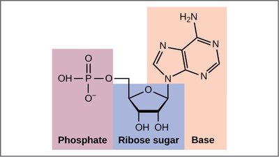
A nucleotide consists of three components: a nitrogenous base, a pentose sugar, and one or more phosphate groups. A nucleoside is composed of only a nitrogenous base and a pentose sugar.
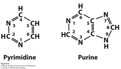
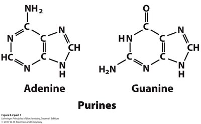
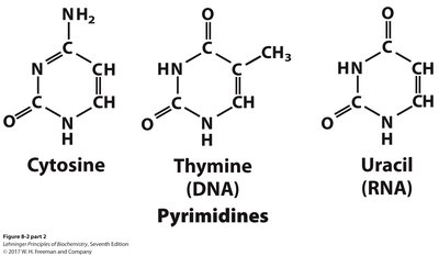
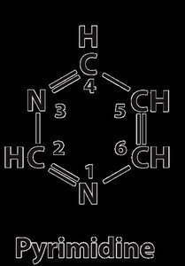
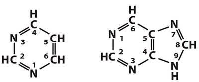
Nitrogenous Base: Purines (adenine, guanine) and pyrimidines (cytosine, thymine, uracil).
Pentose Sugar: Ribose (in RNA) or deoxyribose (in DNA).
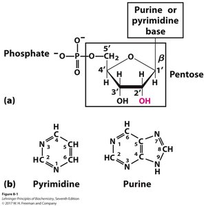
Phosphate Group: Attached to the 5' carbon of the sugar.
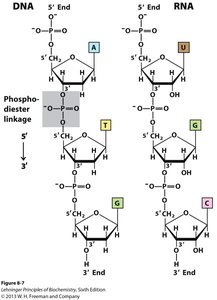
Nucleotide Linkages
Nucleotides are joined by phosphodiester bonds, forming the sugar-phosphate backbone of nucleic acids. The 5' phosphate of one nucleotide connects to the 3' hydroxyl of the next.
Directionality: Nucleic acids have a 5' to 3' direction, crucial for replication and transcription.
Backbone Stability: The phosphodiester bond is resistant to hydrolysis, protecting genetic information.
Pentose Sugars and Their Conformations
Ribose and Deoxyribose
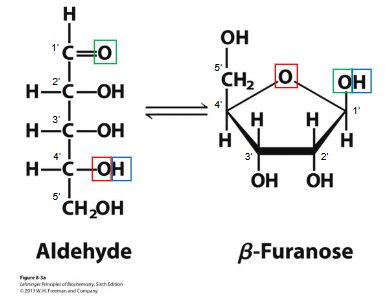
The pentose sugar in nucleotides can exist in straight-chain (aldehyde) or ring (β-furanose) forms. In nucleic acids, the ring form predominates.
Ribose: Found in RNA, contains a 2'-hydroxyl group, making RNA more reactive.
Deoxyribose: Found in DNA, lacks the 2'-hydroxyl group, increasing DNA stability.
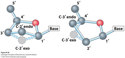
Puckered Conformations
Pentose rings can adopt different puckered conformations, such as C2'-endo and C3'-endo, affecting the overall structure of nucleic acids.
Nitrogenous Bases
Classification and Properties
Nitrogenous bases are classified as purines or pyrimidines. They are planar, aromatic molecules capable of hydrogen bonding and UV absorption.
Purines: Adenine and guanine (found in both DNA and RNA).
Pyrimidines: Cytosine (DNA/RNA), thymine (DNA), uracil (RNA).
Chemical Bonds in DNA and RNA
Types of Bonds
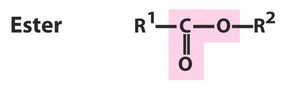
Phosphodiester Bonds: Covalent bonds forming the sugar-phosphate backbone.
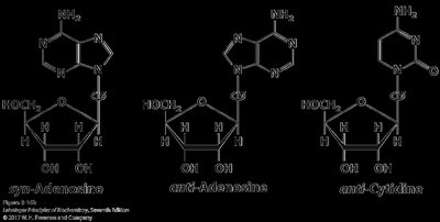
β-N-Glycosidic Bond: Covalent bond linking the base to the sugar (N1 in pyrimidines, N9 in purines).
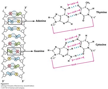
Hydrogen Bonds: Non-covalent bonds between complementary bases (A=T/U, G≡C).
Base Stacking Interactions: Van der Waals and hydrophobic forces between adjacent bases.
Ionic Interactions: Electrostatic interactions with metal ions or proteins.
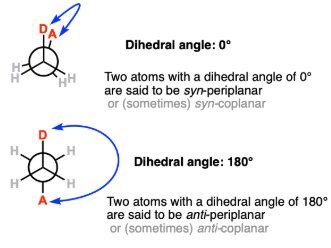
Conformation Around the N-Glycosidic Bond
The N-glycosidic bond allows for syn and anti conformations. The anti conformation is predominant in nucleic acids, facilitating proper base pairing and helix formation.
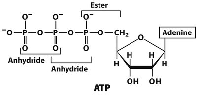
Phosphate Groups and High-Energy Bonds
Phosphate Group Properties
Phosphate groups are negatively charged at physiological pH and are typically attached to the 5' position of the sugar. Nucleic acids are synthesized from nucleoside triphosphates (e.g., ATP, GTP).
High-Energy Phosphoanhydride Bonds
ATP and related nucleotides contain high-energy phosphoanhydride bonds, which release significant energy upon hydrolysis, fueling cellular processes.
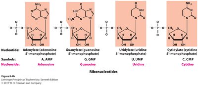
Nomenclature of Nucleotides
Ribonucleotides and Deoxyribonucleotides
Nucleotide names are based on their nitrogenous base, sugar type, and number of phosphate groups. The prefix 'deoxy' is used for DNA nucleotides.
Minor Nucleosides and Modified Bases
DNA Modifications
Minor nucleosides, such as 5-methylcytidine and N6-methyladenosine, are formed post-synthetically and play roles in gene regulation, DNA protection, and repair.
RNA Modifications
Inosine and pseudouridine are common in tRNA and rRNA, contributing to RNA stability and function.
Polynucleotides: Structure and Directionality
Backbone and Sequence
Polynucleotides are linear polymers with a sugar-phosphate backbone and directional 5' and 3' ends. The sequence of bases constitutes the primary structure of DNA or RNA.
Hydrogen Bonding and Base Pairing
Watson-Crick Base Pairs
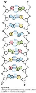
In double-stranded DNA, base pairing occurs between purines and pyrimidines: A pairs with T (2 H-bonds), and G pairs with C (3 H-bonds). This complementarity is essential for DNA replication and transcription.
Historical Discoveries and the Double Helix
Key Milestones
1868: Miescher discovers nuclein (DNA).
1919: Levene identifies nucleotide structure.
1953: Watson and Crick propose the double helix model, aided by Franklin and Wilkins' X-ray data.
Watson-Crick Model
The double helix consists of two antiparallel, complementary strands with a sugar-phosphate backbone on the outside and base pairs inside. Major and minor grooves facilitate protein binding.


DNA Denaturation and Renaturation
Denaturation
Denaturation disrupts secondary structure (hydrogen bonds and base stacking) but leaves the primary structure (covalent bonds) intact. It can be induced by heat or pH changes and is reversible (annealing).
Melting Temperature (Tm): Depends on GC content, DNA length, and ionic strength.
RNA Structure and Function
Types of RNA
mRNA: Carries genetic code from DNA to ribosomes.
tRNA: Brings amino acids to ribosomes during translation.
rRNA: Structural and catalytic component of ribosomes.
miRNA: Regulates gene expression post-transcriptionally.
RNA Secondary Structures
RNA molecules can form complex secondary structures such as hairpins, bulges, internal loops, pseudoknots, and multi-branched loops, which are critical for their function.
Summary Table: Bonds in DNA and RNA
Bond Type | DNA | RNA | Function |
|---|---|---|---|
Phosphodiester Bonds (Covalent) | Present | Present | Forms the sugar-phosphate backbone |
β-N-Glycosidic Bond (Covalent) | Present | Present | Links the nitrogenous base to the sugar |
Hydrogen Bonds (Non-Covalent) | Present (between complementary strands) | Present (in secondary structures) | Stabilizes base pairing |
Base Stacking Interactions | Present | Present (weaker in single-stranded RNA) | Provides additional structural stability |
Ionic Interactions | Present (with backbone and proteins) | Present (with ions and proteins) | Neutralizes negative charge, aids folding |
Conclusion
Nucleotides and nucleic acids are fundamental to life, serving as the blueprint for genetic information and participating in a wide range of cellular processes. Understanding their structure, function, and interactions is essential for the study of biochemistry and molecular biology.
