 Back
BackLEC 17 Oligosaccharides, Polysaccharides, Glycoconjugates, and Glycolysis: Structure, Function, and Metabolic Pathways
Study Guide - Smart Notes
 Tailored notes based on your materials, expanded with key definitions, examples, and context.
Oligosaccharides
Definition and Structure
Oligosaccharides are carbohydrates composed of 2 to 10 monosaccharide units linked by glycosidic bonds. Disaccharides are the simplest oligosaccharides, consisting of two monosaccharide residues. Each monosaccharide unit within an oligosaccharide is referred to as a residue.
Glycosidic bond: The covalent linkage joining two monosaccharides, formed by the reaction of an anomeric carbon with a hydroxyl group of another sugar, resulting in the elimination of water.
Reducing sugar: An oligosaccharide with a free unsubstituted anomeric carbon (e.g., lactose).
Non-reducing sugar: An oligosaccharide without a free anomeric carbon (e.g., sucrose).
Examples: Lactose (milk sugar), cellobiose, sucrose (table sugar), maltose, and isomaltose.

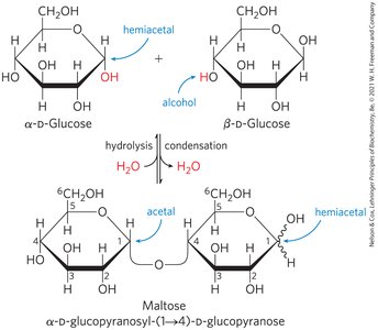
Formation of O-Glycosidic Bonds
O-glycosidic bonds are formed when the hemiacetal or hemiketal group of one monosaccharide reacts with the hydroxyl group of another, producing an acetal or ketal and releasing water. The configuration of the glycosidic bond (α or β) depends on the orientation of the anomeric carbon and the hydroxyl group involved in the linkage.
α-Glycosidic bond: Formed when the hemiacetal and alcohol are on the same side of their rings.
β-Glycosidic bond: Formed when the hemiacetal and alcohol are on opposite sides of their rings.
Polysaccharides
Structure and Function
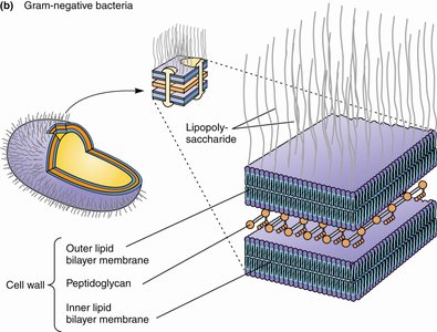
Polysaccharides are large carbohydrates composed of more than 10 monosaccharide units. They serve as storage materials (e.g., starch, glycogen), structural components (e.g., cellulose, peptidoglycan), and protective substances in cells.
Storage polysaccharides: Starch (plants), glycogen (animals)
Structural polysaccharides: Cellulose (plants), peptidoglycan (bacterial cell walls)
Glycoconjugates
Glycoconjugates with Proteins
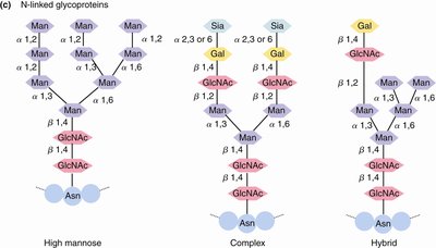
Glycoconjugates are molecules in which carbohydrates are covalently linked to proteins or lipids. Glycoproteins and proteoglycans are important for cell-cell recognition, signaling, and structural integrity.
N-linked glycoproteins: Carbohydrates attached to the nitrogen atom of asparagine side chains.
O-linked glycoproteins: Carbohydrates attached to the oxygen atom of serine or threonine side chains.
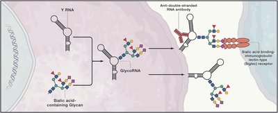
Glycoconjugates with Nucleic Acids
Glycoconjugates can also involve nucleic acids, such as glycoRNAs, which are RNAs modified with carbohydrate groups. These modifications can influence RNA stability, localization, and interactions with proteins.
Glycolysis: The Central Pathway of Glucose Metabolism
Overview and Significance
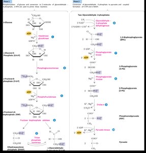
Glycolysis is a universal, anaerobic pathway for the catabolic degradation of glucose to pyruvate, generating ATP and NADH. It is also known as the Embden-Meyerhof pathway and is found in nearly all living cells. Glycolysis consists of ten enzyme-catalyzed reactions, divided into two phases: the energy investment phase and the energy payoff phase.
Phase 1 (Preparatory phase): Phosphorylation of glucose and its conversion to two molecules of glyceraldehyde-3-phosphate. Two ATP molecules are consumed.
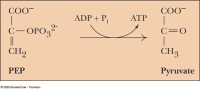
Phase 2 (Payoff phase): Conversion of glyceraldehyde-3-phosphate to pyruvate, producing four ATP and two NADH molecules.
Fates of Pyruvate
The metabolic fate of pyruvate depends on the availability of oxygen and the organism:
Aerobic conditions: Pyruvate is converted to acetyl-CoA, which enters the TCA cycle for complete oxidation to CO2 and H2O.
Anaerobic conditions (muscle): Pyruvate is reduced to lactate (lactic acid fermentation).
Anaerobic conditions (yeast): Pyruvate is converted to ethanol and CO2 (alcoholic fermentation).
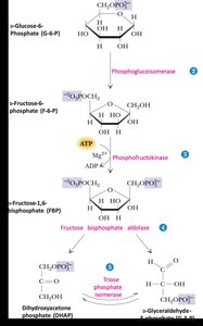
Glycolysis: Phase 1 – Preparatory Reactions
Reaction 1: Phosphorylation of Glucose
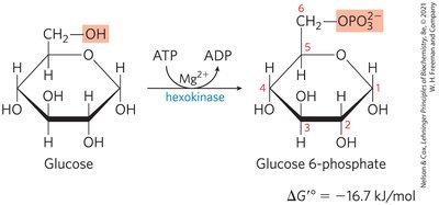
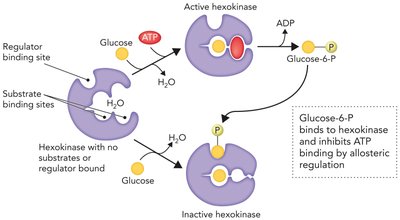
The first step of glycolysis is the phosphorylation of glucose to glucose-6-phosphate, catalyzed by hexokinase (or glucokinase in the liver and pancreas). This reaction consumes one ATP molecule and is highly exergonic, helping to trap glucose inside the cell and regulate glycolytic flux.
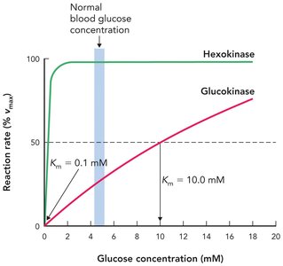
Enzyme: Hexokinase (low Km, high affinity; inhibited by glucose-6-phosphate) or glucokinase (high Km, low affinity; not inhibited by glucose-6-phosphate, induced by insulin).
Equation:
Standard free energy change:
Physiological Importance of Glucose Phosphorylation
Phosphorylation adds a negative charge, preventing glucose-6-phosphate from crossing the plasma membrane by passive diffusion.
Maintains a low intracellular glucose concentration, favoring continued glucose uptake by facilitated diffusion.
Phosphorylated intermediates are essential for substrate-level phosphorylation and energy yield in glycolysis.
Hexokinase vs. Glucokinase
Hexokinase: Expressed in most tissues, low Km (0.1–0.3 mM), inhibited by glucose-6-phosphate.
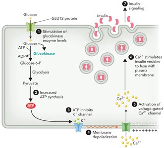
Glucokinase: Expressed in liver and pancreas, high Km (5–10 mM), not inhibited by glucose-6-phosphate, induced by insulin.
In diabetes, low insulin leads to reduced glucokinase and glycogen synthesis in the liver, resulting in high blood glucose.
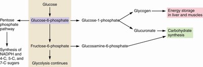
Glucose-6-Phosphate as a Metabolic Hub
Glucose-6-phosphate is a central intermediate that can enter multiple metabolic pathways, including glycolysis, glycogen synthesis, the pentose phosphate pathway, and carbohydrate biosynthesis.
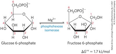
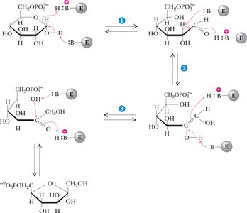
Reaction 2: Phosphoglucoisomerase
Phosphoglucoisomerase (also called phosphohexose isomerase) catalyzes the reversible isomerization of glucose-6-phosphate (an aldose) to fructose-6-phosphate (a ketose). This reaction proceeds via an enediol intermediate and requires Mg2+ as a cofactor.
Equation:
Standard free energy change:
Operates near equilibrium and is readily reversible.
Physiological Rationale for Isomerization
Phosphorylation at C-1 is more efficient with a primary alcohol (fructose-6-phosphate) than with a hemiacetal (glucose-6-phosphate).
Isomerization activates the bond between C-3 and C-4 for cleavage in the subsequent aldolase reaction.
Recap Questions and Answers
How is glucose kept inside the cell, against a concentration gradient? Answer: By conversion to glucose-6-phosphate. Phosphorylation traps glucose inside the cell because the phosphate group has a negative charge at physiological pH.
Which carbons of the original glucose are phosphorylated in the two molecules of glyceraldehyde 3-phosphate? Answer: C-1 and C-6. The first three glycolytic reactions convert glucose to fructose 1,6-bisphosphate. After cleavage by aldolase, C-1 becomes the phosphorylated carbon of dihydroxyacetone phosphate and C-6 becomes the phosphorylated carbon of glyceraldehyde 3-phosphate. Triose phosphate isomerase converts dihydroxyacetone phosphate to the second glyceraldehyde 3-phosphate molecule.
