 Back
BackProtein Folding: Mechanisms, Chaperones, and Disease
Study Guide - Smart Notes
 Tailored notes based on your materials, expanded with key definitions, examples, and context.
Protein Folding
Introduction to Protein Folding
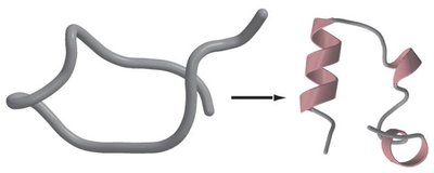
Protein folding is the process by which a polypeptide chain acquires its biologically functional three-dimensional structure. The sequence of amino acids in a protein dictates its final conformation, which is essential for its function. Understanding protein folding is crucial for predicting protein structure and for applications in protein engineering and biotechnology.
Protein folding problem: Refers to the challenge of predicting a protein's native structure from its amino acid sequence.
Significance: Proper folding is necessary for protein function; misfolding can lead to disease.
Applications: Industrial production of proteins often requires refolding from denatured states.
Mechanisms and Pathways of Protein Folding
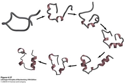
Protein folding is a thermodynamically driven process that occurs through a series of intermediate states. The process is not random but follows specific pathways that minimize the free energy of the system.
Thermodynamics: Folding is favored when the overall free energy change () is negative.
Key factors:
Conformational entropy (unfavorable, opposes folding)
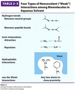
Non-covalent interactions (hydrogen bonds, ionic interactions, van der Waals forces, hydrophobic effect)
Disulfide bonds (covalent stabilization)-A covalent bond is a chemical bond formed when two atoms share pairs of valence electrons in order to achieve stability and a full outer electron shell.
Folding intermediates: Proteins often pass through partially folded states, such as the "molten globule" state, before reaching their native conformation.
Thermodynamics of Protein Folding
The folding of globular proteins is governed by a balance of enthalpic and entropic contributions. The process is spontaneous under physiological conditions due to the following:
Unfavorable conformational entropy: Loss of randomness as the protein adopts a specific structure.
Favorable enthalpy: Formation of intramolecular interactions (hydrogen bonds, ionic interactions).
Favorable entropy (hydrophobic effect): Burying hydrophobic residues increases the entropy of water molecules.
The overall free energy change is given by:
Stabilizing Interactions in Proteins
Protein stability is achieved through a combination of non-covalent and covalent interactions:
Electrostatic interactions: Ionic bonds and salt bridges between charged side chains.
Hydrogen bonds: Between backbone and side chain groups.
Van der Waals interactions: Weak attractions between all atoms in close proximity.
Hydrophobic interactions: Nonpolar side chains cluster away from water.
Disulfide bonds: Covalent bonds between cysteine residues stabilize the folded structure.
Denaturation and Misfolding
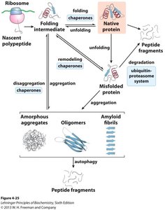
Proteins are sensitive to their environment. Changes in temperature, pH, or the presence of chemicals can disrupt their structure, leading to denaturation—a loss of function due to structural changes. Misfolded proteins can aggregate and are often targeted for degradation, but persistent misfolding can cause disease.
Denaturation: Partial or complete unfolding, often reversible.
Misfolding: Incorrect folding can lead to aggregation and disease.

Assisted Protein Folding: Molecular Chaperones and Chaperonins
Molecular Chaperones
Not all proteins fold spontaneously. Molecular chaperones are specialized proteins that assist in the folding of other proteins, preventing aggregation and misfolding. They do not form part of the final structure but facilitate correct folding.
Hsp70 family: Includes Hsp70 (DnaK in bacteria), Hsp40 (DnaJ), and GrpE. These proteins bind to hydrophobic regions of unfolded polypeptides, preventing aggregation and assisting in folding through ATP-dependent cycles.
Functions:
Protect nascent or stress-denatured proteins
Block premature folding during translocation
Facilitate refolding or degradation of misfolded proteins

Chaperonins: GroEL and GroES
Chaperonins are large, multi-subunit complexes that provide an isolated environment for protein folding. In Escherichia coli, GroEL and GroES are the primary chaperonins, essential for the folding of many cellular proteins, especially under stress conditions.
GroEL: Forms a double-ring structure with a central cavity for protein folding.
GroES: Acts as a cap, enclosing the substrate protein within the GroEL chamber.
Mechanism: Unfolded proteins bind GroEL, GroES caps the complex, and ATP hydrolysis drives conformational changes that promote folding.

Other Folding Assistants
Protein disulfide isomerase (PDI): Catalyzes the formation and rearrangement of disulfide bonds.
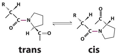
Peptide prolyl cis-trans isomerase (PPI): Catalyzes the interconversion of cis and trans isomers of proline peptide bonds, facilitating proper folding.
Protein Misfolding and Disease
Consequences of Protein Misfolding
Misfolded proteins can be degraded or may aggregate, leading to cellular dysfunction and disease. A significant fraction of newly synthesized proteins are degraded due to misfolding.
Sickle-cell anemia: Caused by a single amino acid substitution (Glu6 to Val6) in hemoglobin, leading to aggregation and fiber formation.
Amyloidoses: Diseases caused by the accumulation of insoluble amyloid fibrils, which are highly ordered aggregates rich in β-sheet structure.
 ;
;
Examples of Protein Misfolding Diseases
Alzheimer’s disease: Aggregation of Tau protein into amyloid-like fibrils.
Huntington’s disease: Aggregation of Huntingtin protein due to polyglutamine repeats.
Parkinson’s disease: Aggregation of α-synuclein into filamentous masses.
Cystic fibrosis: Misfolding of the CFTR chloride channel due to a deletion mutation.
Prions and Infectious Protein Misfolding
Prions are infectious proteins that propagate by inducing misfolding of normal cellular proteins. Prion diseases are fatal neurodegenerative disorders characterized by spongiform changes in brain tissue.
Examples: Mad cow disease (BSE), Creutzfeldt-Jakob disease, kuru, scrapie, chronic wasting disease.
Mechanism: The normal prion protein (PrP) is converted to a misfolded, disease-causing form (PrPSc), which induces further misfolding in a self-propagating manner.
Summary Table: Key Factors in Protein Folding
Factor | Role in Folding |
|---|---|
Conformational Entropy | Opposes folding (loss of randomness) |
Hydrophobic Effect | Favors folding (burial of nonpolar residues) |
Internal Interactions | Favors folding (hydrogen bonds, ionic, van der Waals) |
Disulfide Bonds | Stabilize folded structure |
Molecular Chaperones | Assist folding, prevent aggregation |
Chaperonins | Provide isolated folding environment |
