 Back
Protein Folding, Stability, and Fibrous Proteins: Structure and Function
Study Guide - Smart Notes
 Tailored notes based on your materials, expanded with key definitions, examples, and context.
Protein Folding and Tertiary Structure
Anfinsen’s Renaturation Experiments
Christian Anfinsen’s classic experiments with ribonuclease A demonstrated that proteins can spontaneously refold into their native, functional conformations under physiological conditions. These experiments provided foundational evidence that the information required for proper protein folding is encoded entirely within the primary amino acid sequence.
Denaturation: Treatment with 8M urea and mercaptoethanol unfolds the protein and reduces disulfide bonds.
Renaturation: Removal of denaturants allows the protein to refold and reform correct disulfide bonds, restoring activity.
Scrambled Conformation: If disulfides reform before proper folding, the protein may become trapped in an inactive, scrambled state.
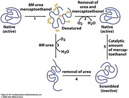
Additional info: These findings led to Anfinsen’s postulate: the native structure of a protein is the thermodynamically most stable conformation possible for its sequence.
How Do Proteins Fold?
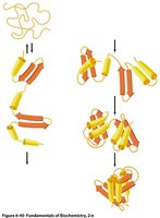
Protein folding is a complex, yet highly efficient process that typically occurs in two phases:
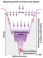
Fast Phase: Local secondary structures form rapidly, often driven by local sequence preferences, leading to a hydrophobic collapse into a "molten globule" state.
Slow Phase: Secondary structures stabilize, side chains pack, and water is expelled from the core, resulting in the native tertiary structure.
Amphipathic Secondary Structure
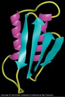
Amphipathic structures contain both hydrophobic and hydrophilic regions. In β-sheets, alternating polar and nonpolar side chains create distinct faces, influencing their location within the protein or at interfaces such as membranes. Amphipathic α-helices can be predicted using helical wheel diagrams, which reveal the distribution of polar and nonpolar residues around the helix.
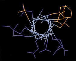
Additional info: Buried helices tend to be more hydrophobic, while surface helices are more amphipathic.
The Folding Funnel Model
The folding funnel describes the energy landscape of protein folding. Proteins fold through a series of conformational changes that progressively decrease free energy and entropy, eventually reaching the native state. There are many possible folding pathways, and proteins may transiently become trapped in local minima.
Driving Forces in Protein Folding
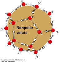
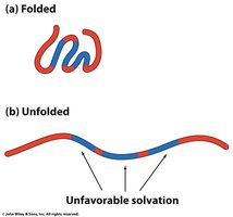
The primary driving force for protein folding is the hydrophobic effect, which is entropically driven. Hydrophobic residues cluster in the protein core, minimizing their exposure to water and increasing the entropy of the surrounding solvent.
Hydrogen Bonds: Important for defining structure, but not the main contributor to stability due to desolvation costs.
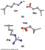
Ion Pairs/Salt Bridges: Can contribute to specificity and stability, especially when buried, but are less significant on the protein surface due to competition with water.
Protein Denaturation
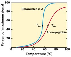
Denaturation refers to the loss of secondary and tertiary structure, resulting in a random coil. The temperature at which half the protein population is unfolded is called the melting temperature (Tm). Denaturation can be induced by heat, chemicals, or extreme pH, and is often a cooperative process.
Protein Misfolding and Disease
Protein Misfolding
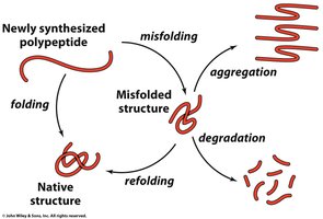
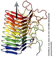
Proteins can misfold, leading to aggregation or degradation. Misfolded proteins are implicated in several diseases, including Alzheimer’s, Huntington’s, and prion diseases. Prion proteins can exist in a normal (PrPC) or infectious (PrPSc) form, with the latter catalyzing the conversion of the former, leading to disease.
Molecular Chaperones
Role of Chaperones
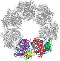
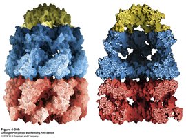
Molecular chaperones assist in protein folding by preventing inappropriate interactions and aggregation. Chaperonins, such as the GroEL/GroES complex, provide an isolated environment for proper folding, using ATP hydrolysis to drive conformational changes.
Fibrous Proteins: Structure and Function
Overview of Fibrous Proteins
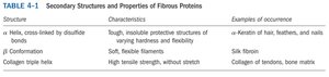
Fibrous proteins are characterized by repetitive secondary structures and serve primarily structural roles in organisms. They are generally insoluble and form the basis of materials such as hair, silk, and connective tissue.
Structure | Characteristics | Examples of Occurrence |
|---|---|---|
α Helix, cross-linked by disulfide bonds | Rough, insoluble protective structures of varying hardness and flexibility | Keratin of hair, feathers, and nails |
β Conformation | Soft, flexible filaments | Silk fibroin |
Collagen triple helix | High tensile strength, without stretch | Collagen of tendons, bone matrix |
Keratin
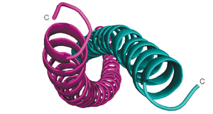
Keratin is a fibrous protein forming coiled-coil α-helices stabilized by hydrophobic interactions and disulfide bonds. It is the main component of hair, nails, and animal claws. The quaternary structure involves multiple helices cross-linked for strength and elasticity.
Silk Fibroin
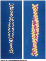
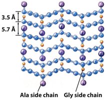
Silk fibroin consists of extended β-sheets with repeating Gly-Ala sequences. This structure provides flexibility but little stretch, as the sheets are already fully extended.

Collagen
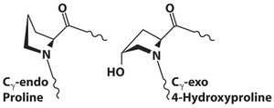
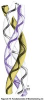
Collagen is the most abundant protein in connective tissues. It is composed of three left-handed helices twisted into a right-handed triple helix. The sequence is rich in glycine, proline, and hydroxyproline, with post-translational modifications critical for stability.
Gly-X-Y motif: X is often proline, Y is often hydroxyproline.
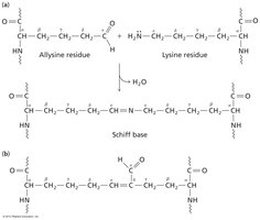
Crosslinking: Covalent bonds between modified lysine residues (allysine) further stabilize the structure.
Collagen and Scurvy
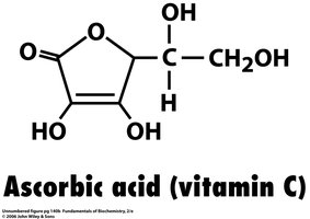
Vitamin C (ascorbic acid) is essential for the enzymatic conversion of proline to hydroxyproline in collagen biosynthesis. Deficiency leads to scurvy, a disease characterized by weakened connective tissue. This can be reversed by restoring vitamin C intake.
Key Concepts and Applications
Protein folding is a two-step process driven primarily by the hydrophobic effect.
Chaperonins assist in proper folding and prevent aggregation.
Fibrous proteins have specialized structures for mechanical support and protection.
Post-translational modifications are critical for the function and stability of structural proteins like collagen.
