 Back
BackProtein Function and Evolution: Analytical Methods, Antibody Structure, and Hemoglobin Allostery
Study Guide - Smart Notes
 Tailored notes based on your materials, expanded with key definitions, examples, and context.
Protein Analysis Techniques
Polyacrylamide Gel Electrophoresis (PAGE)
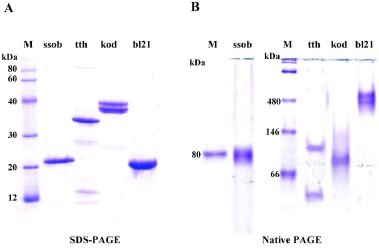
Polyacrylamide gel electrophoresis (PAGE) is a fundamental technique for separating proteins based on their size and charge. There are several variations of PAGE, each with specific applications in protein analysis.
Native PAGE: Proteins are separated in their native, folded state, preserving their structure and function. This method allows for the analysis of protein complexes and oligomeric states.
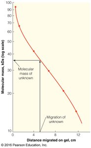
Denaturing PAGE (SDS-PAGE): Proteins are denatured using sodium dodecyl sulfate (SDS), which imparts a uniform negative charge and linearizes the proteins. Separation is based solely on molecular weight.
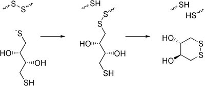
Reducing vs. Non-Reducing PAGE: Reducing agents such as dithiothreitol (DTT) or β-mercaptoethanol (BME) can be added to break disulfide bonds, allowing for the analysis of subunit composition.
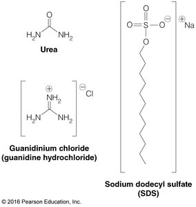
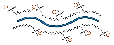
Key Point: In SDS-PAGE, the migration of proteins depends only on their length (molecular weight), as SDS masks intrinsic charges and shapes.
Reducing Agents: DTT and BME are commonly used to reduce disulfide bonds in proteins, facilitating the analysis of subunit composition.

Example: SDS-PAGE can distinguish between the heavy and light chains of antibodies under reducing conditions.
Blotting Techniques
Western, Southern, and Northern Blots
Blotting techniques are used to detect specific biomolecules after electrophoretic separation:
Western Blot: Detects specific proteins using antibodies after transfer to a membrane.
Southern Blot: Detects specific DNA sequences using labeled DNA probes.
Northern Blot: Detects specific RNA sequences using labeled DNA probes.
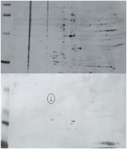
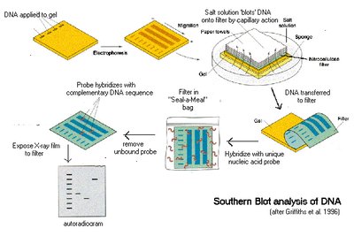
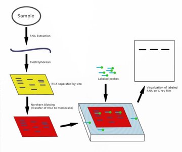
Example: Western blots can identify phosphorylated proteins in a complex mixture using phospho-specific antibodies.
Antibody Structure and Function
Antigen Recognition and Immune Response
Antibodies (immunoglobulins) are proteins produced by B cells that specifically recognize antigens. Each antibody binds to a unique epitope on the antigen, initiating an adaptive immune response.
Antigen: A molecule that elicits an immune response.
Epitope: The specific part of the antigen recognized by the antibody.
Antibody: A Y-shaped protein that binds antigens with high specificity.
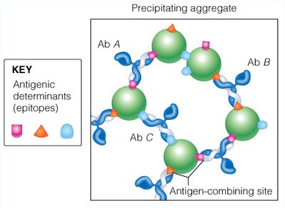
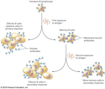
Example: Vaccines stimulate the production of memory B cells, providing long-term immunity.
Antibody Structure
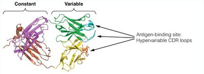
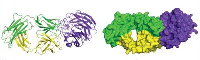
Antibodies are composed of two heavy and two light chains, forming a Y-shaped molecule. The variable regions at the tips of the Y form the antigen-binding sites, which are highly specific due to hypervariable complementarity-determining regions (CDRs).
Fab Fragment: Contains the antigen-binding site.
Fc Fragment: Mediates effector functions but does not bind antigen.
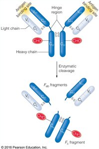
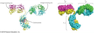
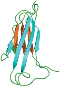
Key Point: Antibody-antigen interactions are mediated by shape and charge complementarity, involving hydrogen bonds, van der Waals forces, and electrostatic interactions.
Hemoglobin and Myoglobin: Structure and Function
Oxygen Transport Proteins
Hemoglobin and myoglobin are globular proteins that bind oxygen via a heme prosthetic group. Myoglobin is a monomer found in muscle, while hemoglobin is a tetramer found in red blood cells.
Myoglobin (Mb): Stores O2 in muscle tissue for use during periods of high demand.
Hemoglobin (Hb): Transports O2 from lungs to tissues and returns CO2 for exhalation.

Key Point: Both proteins share a common globin fold and bind heme, but differ in quaternary structure and oxygen-binding properties.
Heme and Oxygen Binding
The heme group is a conjugated tetrapyrrole ring (protoporphyrin IX) with a central Fe2+ ion. Oxygen binds reversibly to the Fe2+ ion, which is coordinated by four nitrogen atoms of the porphyrin ring, a proximal histidine, and O2.
Oxyhemoglobin/Oxymyoglobin: Oxygen-bound forms.
Deoxyhemoglobin/Deoxymyoglobin: Oxygen-free forms.
Oxygen Binding Curves and P50
Oxygen binding to myoglobin follows a hyperbolic curve, described by the equation:
P50: The partial pressure of O2 at which half of the binding sites are occupied; a measure of affinity.
A lower P50 indicates higher affinity for O2.
Allosteric Regulation of Hemoglobin
Hemoglobin exhibits cooperative binding, resulting in a sigmoidal O2 binding curve. This is explained by allosteric transitions between the T (tense, low affinity) and R (relaxed, high affinity) states.
Homotropic Allostery: O2 is a positive homotropic effector; its binding increases affinity at other sites.
Heterotropic Allostery: H+, CO2, and 2,3-BPG are negative effectors; their binding decreases O2 affinity.
The Hill equation describes cooperative binding:
where n is the Hill coefficient (degree of cooperativity).
Mechanisms of Allostery: MWC and KNF Models
MWC (Monod-Wyman-Changeux) Model: Hemoglobin exists in equilibrium between T and R states; O2 binding shifts equilibrium toward R.
KNF (Koshland-Némethy-Filmer) Model: O2 binding induces sequential conformational changes in subunits.
Allosteric Effectors and the Bohr Effect
Allosteric effectors modulate hemoglobin's affinity for oxygen:
H+ (Bohr Effect): Lower pH (higher H+) stabilizes the T state, promoting O2 release.
CO2: Binds to hemoglobin and forms carbamates, stabilizing the T state and contributing to the Bohr effect.
2,3-Bisphosphoglycerate (2,3-BPG): Binds in the central cleft of deoxyhemoglobin, stabilizing the T state and facilitating O2 release in tissues.
Example: Fetal hemoglobin has reduced BPG binding, resulting in higher O2 affinity for efficient transfer from maternal blood.
Protein Evolution and Hemoglobinopathies
Evolutionary Relationships
Gene duplication and mutation drive the evolution of globin proteins. Sequence comparisons reveal evolutionary trees and functional divergence among globins.
Hemoglobinopathies: Genetic disorders such as sickle-cell disease arise from mutations in globin genes, affecting protein function and red blood cell morphology.
Sickle-Cell Disease: A single amino acid substitution leads to hemoglobin polymerization, red cell deformation, and anemia. Heterozygotes are resistant to malaria.
Summary Table: Key Differences Between Myoglobin and Hemoglobin
Feature | Myoglobin | Hemoglobin |
|---|---|---|
Structure | Monomer | Tetramer (2α, 2β) |
Location | Muscle tissue | Red blood cells |
O2 Binding Curve | Hyperbolic | Sigmoidal (cooperative) |
Function | O2 storage | O2 transport |
Allosteric Regulation | None | Yes (H+, CO2, 2,3-BPG) |
