 Back
Protein Function and Evolution: Structure, Analysis, and Allosteric Regulation
Study Guide - Smart Notes
 Tailored notes based on your materials, expanded with key definitions, examples, and context.
Protein Function and Evolution
Polyacrylamide Gel Electrophoresis (PAGE)
Polyacrylamide gel electrophoresis (PAGE) is a fundamental technique used to separate proteins and nucleic acids based on their size, charge, and structural state. It is essential for analyzing protein purity, molecular weight, and structural integrity.
Native Gel: Preserves the native structure of molecules, allowing analysis of protein complexes and functional states.
Denaturing Gel: Uses agents like SDS (for proteins) or urea (for nucleic acids) to destroy native structure, enabling separation solely by size.
Reducing Gel: Includes reducing agents (e.g., DTT, β-mercaptoethanol) to break disulfide bonds, further denaturing proteins.
Non-reducing Gel: Preserves disulfide bonds, useful for studying quaternary structure.
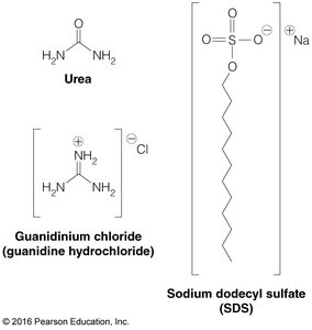
SDS-PAGE is a denaturing gel technique where SDS coats proteins, imparting a uniform negative charge and allowing separation by molecular weight alone.
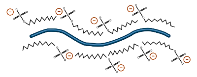
Protein shape and native charge are irrelevant; migration depends only on peptide length (molecular weight).
Reducing agents such as DTT and β-mercaptoethanol break disulfide bonds, enabling analysis of subunit composition.
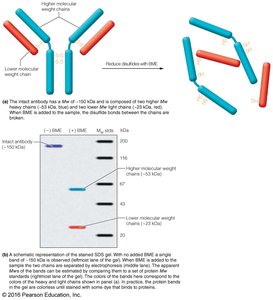

Comparison of SDS-PAGE and native PAGE reveals differences in protein migration due to structural preservation or denaturation.
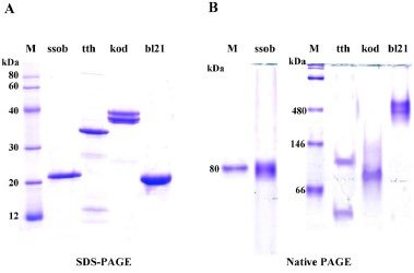
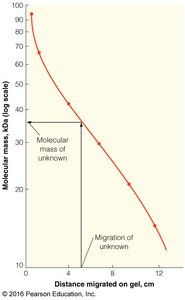
The migration distance in SDS-PAGE is inversely related to molecular mass, allowing estimation of unknown protein sizes.
Antibody Structure and Function
Antibodies are immunoglobulins that recognize specific epitopes on antigens, forming the basis of the adaptive immune response.
Antigen: Substance that elicits an immune response.
Antibody: Protein that binds specifically to an antigen.
Epitope: Region of antigen recognized by antibody.
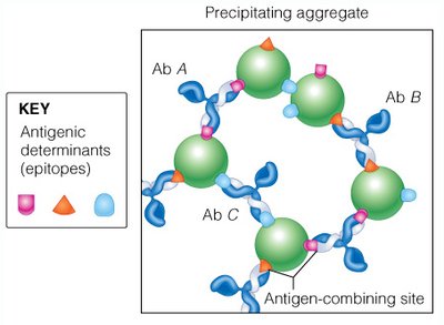
Antibodies can crosslink antigens, forming precipitating aggregates.
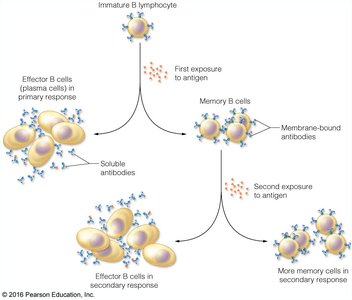
B-lymphocytes display antibodies and, upon antigen exposure, differentiate into effector and memory cells, providing the basis for vaccination.
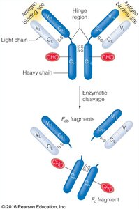
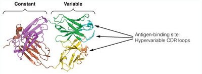
IgG antibodies consist of two heavy and two light chains, with enzymatic cleavage at the hinge region producing Fab (antigen-binding) and Fc (constant) fragments.
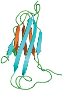
The immunoglobulin fold is a β-sandwich motif, composed of two antiparallel β sheets, occurring multiple times in IgG molecules.
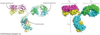
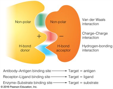
Antibody-antigen interactions are mediated by shape and charge complementarity, primarily at hypervariable CDR loops.
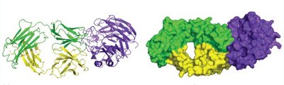
These interactions involve hydrogen bonds, salt bridges, and hydrophobic contacts.
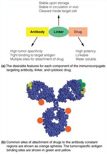
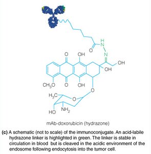
Antibodies are used in targeted drug delivery due to their high specificity.
Blotting Techniques
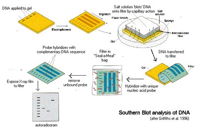
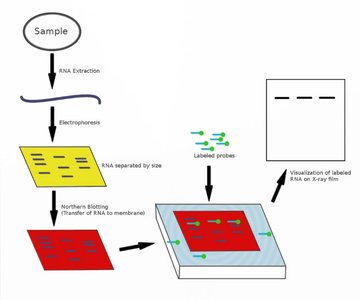
Blotting techniques are used to detect specific biomolecules in complex mixtures:
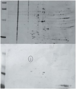
Western Blot: Detects proteins using antibodies after electrophoresis and transfer to a membrane.
Southern Blot: Detects DNA sequences using radioactive DNA probes.
Northern Blot: Detects RNA sequences using radioactive DNA probes.
Hemoglobin and Myoglobin: Structure and Function
Globins are heme proteins involved in oxygen transport and storage. Myoglobin is a monomeric protein found in muscle, while hemoglobin is a tetrameric protein in red blood cells.
Myoglobin: Efficiently delivers O2 to mitochondria; abundant in deep-diving mammals.
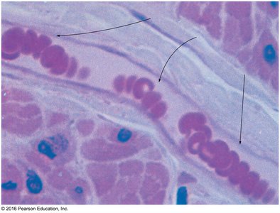
Hemoglobin: Transports O2 from lungs/gills to tissues and returns CO2 for exhalation.
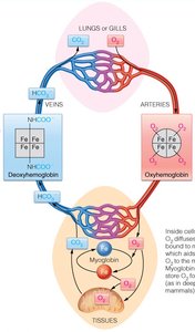


Both proteins share a common folding motif and contain a heme group, an iron porphyrin.
Apoprotein: Protein without heme.
Holoprotein: Protein with heme.
Oxyhemoglobin/Oxymyoglobin: O2-bound forms.
Deoxyhemoglobin/Deoxymyoglobin: O2-free forms.
O2 binds at the Fe2+ center of heme, coordinated by four N atoms, one O2, and the proximal histidine residue. The distal histidine stabilizes O2 via hydrogen bonding. CO binds more strongly than O2.
O2 binding can be monitored by color change, explaining the color difference between veins and arteries.
Oxygen Binding Curves and P50
The oxygen binding curve for myoglobin is hyperbolic, described by the following equations:
P50 is the partial pressure of O2 at half saturation; lower P50 indicates higher affinity.
Hemoglobin displays a sigmoidal binding curve, reflecting cooperative binding and allosteric regulation.
(non-cooperative)
Hill equation:
Allosteric Regulation of Hemoglobin
Allostery refers to the regulation of protein activity by binding of effectors at sites other than the active site. Hemoglobin exhibits both homotropic (O2) and heterotropic (H+, CO2, BPG) allosteric regulation.
Homotropic Effectors: Bind at the active site (e.g., O2).
Heterotropic Effectors: Bind at other sites (e.g., H+, CO2, 2,3-BPG).
Positive Allostery: Increases O2 affinity (O2 binding).
Negative Allostery: Decreases O2 affinity (H+, CO2, BPG binding).
Hemoglobin transitions between T (tense, low O2 affinity) and R (relaxed, high O2 affinity) states. The KNF and MWC models describe these transitions:
KNF Model: O2 binding induces conformational change in one subunit, facilitating changes in adjacent subunits.
MWC Model: Hemoglobin exists in T or R states; O2 binding shifts equilibrium toward R.
O2 binding at heme induces conformational changes via the Perutz mechanism, involving movement of Fe2+ and proximal histidine.
Allosteric Effectors of Hemoglobin
Allosteric effectors modulate hemoglobin's O2 binding affinity:
H+ (Bohr Effect): Low pH favors T state, increasing O2 delivery.
CO2: Binds as carbamate, stabilizing T state and contributing to Bohr effect.
2,3-BPG: Binds in T state cleft, lowering O2 affinity and promoting O2 release.
Fetal hemoglobin has reduced BPG binding, favoring R state and higher O2 affinity.
Protein Evolution and Hemoglobinopathies
Mutations in globin genes define evolutionary relationships and can cause hemoglobinopathies such as sickle-cell disease. Sickle-cell disease results from abnormal erythrocytes, leading to anemia and increased malaria resistance in heterozygotes.
Summary Table: Comparison of Myoglobin and Hemoglobin
Property | Myoglobin | Hemoglobin |
|---|---|---|
Structure | Monomeric | Tetrameric (α2β2) |
Function | O2 storage in muscle | O2 transport in blood |
O2 Binding Curve | Hyperbolic | Sigmoidal |
Allosteric Regulation | None | Yes (T/R states, effectors) |
P50 | Low (high affinity) | Variable (regulated) |
Additional info: Academic context was added to clarify mechanisms, equations, and structural details for completeness and exam preparation.
