 Back
Lecture 7 - Ligand Binding
Study Guide - Smart Notes
 Tailored notes based on your materials, expanded with key definitions, examples, and context.
Protein Function and Ligand Binding
General Description of Ligand Binding
Proteins often function by binding to specific molecules called ligands. Ligands can include small molecules, ions, DNA/RNA, or other proteins. The binding is typically reversible and non-covalent, allowing proteins to respond dynamically to changes in their environment.
Ligand: A molecule that reversibly binds to a protein, affecting its activity or function.
Apo form: The state of a protein without a bound ligand.
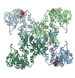
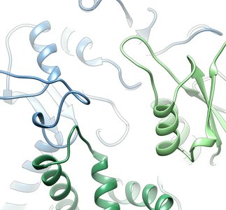
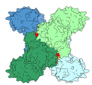
Conformational changes: Ligand binding can induce structural changes in proteins, altering their activity (e.g., switching between "on" and "off" states).
Protein Structures Are Not Static
Proteins exhibit flexibility, and their structure can change depending on ligand binding, post-translational modifications (PTMs), or interactions with other partners. These conformational changes are essential for protein function.
Rapidly reversible binding: Ligand binding and release can occur quickly, allowing proteins to adapt to cellular needs.
Functional diversity: Different conformations correspond to different functional states.
Quantitative Description of Ligand Binding
Binding Equilibrium and Dissociation Constant (Kd)
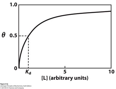
The equilibrium between a protein (P) and ligand (L) is described by the dissociation constant (Kd), which quantifies the affinity of the protein for its ligand.
Binding reaction: P + L → P•L
Dissociation constant:
Units: Concentration (typically Molar, M)
Fractional occupancy (Θ):
Interpretation of Kd
The value of Kd indicates the affinity of the protein for its ligand:
Smaller Kd: Higher affinity, tighter binding
Larger Kd: Lower affinity, weaker binding
At [L] = Kd: Half of the binding sites are occupied ()
Oxygen Binding Proteins: Myoglobin and Hemoglobin
Myoglobin: Hyperbolic O2 Binding
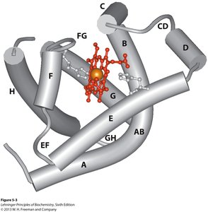
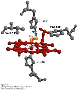
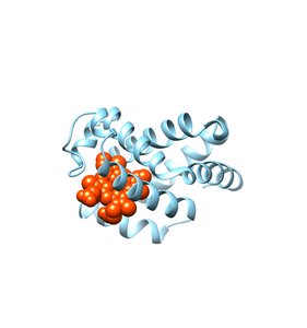
Myoglobin (Mb) is a single polypeptide chain with a heme group that binds oxygen. It serves as an oxygen storage protein, especially in muscle tissue.
Structure: 1 polypeptide, 8 α-helices, 1 heme group
O2 binding: O2 binds to Fe in heme and to HisE7 in the protein
Binding equation:
p50: Partial pressure of O2 at which half of the sites are occupied
Example: Myoglobin Oxygen Affinity
p50 for Mb: 0.26 kPa
In lungs (pO2 = 13.3 kPa): (98% occupied)
In tissue (pO2 = 4 kPa): (94% occupied)
Hemoglobin: Sigmoidal O2 Binding and Cooperativity
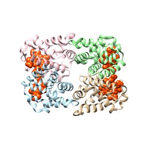
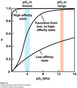
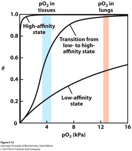
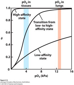
Hemoglobin (Hb) is a tetrameric protein responsible for oxygen transport from lungs to tissues. Its O2 binding curve is sigmoidal, reflecting cooperative binding among its four subunits.
Structure: 4 polypeptide chains, each with a heme group
Function: Delivers O2 efficiently by releasing it in tissues
Cooperativity: Binding of O2 to one subunit increases the affinity of the remaining subunits
Sigmoidal curve: Steep transition between low and high affinity states
Example: Hemoglobin Oxygen Release
In lungs (pO2 = 13.3 kPa):
In tissue (pO2 = 4 kPa):
O2 released: (40% of O2 released)
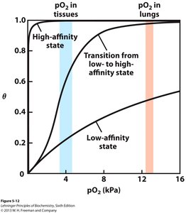
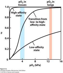
Mechanism of Cooperativity: T and R States
Hemoglobin can exist in two main conformational states:
T (tense) state: Low affinity for O2
R (relaxed) state: High affinity for O2
Transition: Increasing pO2 shifts equilibrium from T to R state
Positive cooperativity: Binding at one site increases affinity at other sites
General Expression for Multiple Ligand Binding
For proteins with multiple binding sites, the binding curve can be described by the Hill equation:
Hill equation:
N: Number of binding sites
n: Hill coefficient (degree of cooperativity)
As n increases: The curve becomes more sigmoidal (steeper transition)
Limiting Cases
N=1, n=1: Hyperbolic binding (e.g., myoglobin)
N>1, n=1: Independent binding sites
N>1, n=N: Maximum cooperativity (steepest curve)
Summary Table: Myo1globin vs. Hemoglobin
Protein | Structure | O2 Binding Curve | Function | Cooperativity |
|---|---|---|---|---|
Myoglobin | Monomer, 1 heme | Hyperbolic | O2 storage | None |
Hemoglobin | Tetramer, 4 hemes | Sigmoidal | O2 transport | Positive |
Key Concepts for Exam Preparation
Understand the definition and role of ligands in protein function
Be able to use the general expression for ligand binding to interpret binding curves and predict fractional occupancy
Recognize the difference between hyperbolic (myoglobin) and sigmoidal (hemoglobin) binding curves
Explain the concept of cooperative binding and its physiological significance
Apply the Hill equation to describe binding behavior in proteins with multiple sites
Additional info: The notes expand on the original content by providing definitions, equations, and examples for clarity and completeness. All included images directly reinforce the explanation of protein structure, ligand binding, and cooperativity.
