 Back
Protein Structure: Hierarchical Organization and Peptide Bond Chemistry
Study Guide - Smart Notes
 Tailored notes based on your materials, expanded with key definitions, examples, and context.
Protein Structure
Levels of Protein Structure
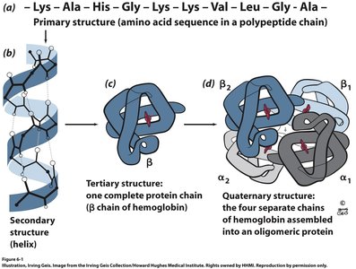
Proteins exhibit a hierarchical structure, which is essential for their biological function. The four levels of protein structure are primary, secondary, tertiary, and quaternary.
Primary Structure: The linear sequence of amino acids in a polypeptide chain, determined by the genetic code. Each amino acid is connected by a peptide bond, and the sequence dictates the protein's folding and function.
Secondary Structure: Localized folding patterns stabilized by hydrogen bonds, primarily forming α-helices and β-sheets.
Tertiary Structure: The overall three-dimensional shape of a single polypeptide chain, resulting from interactions among side chains (R groups).
Quaternary Structure: The assembly of multiple polypeptide chains (subunits) into a functional protein complex.
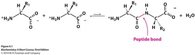
Peptide Bond Formation
Peptide bonds are formed via a condensation reaction between the amino group of one amino acid and the carboxyl group of another, releasing water. This bond is fundamental to the primary structure of proteins.
Condensation Reaction:
Peptide Bond: The covalent bond linking the carbonyl carbon of one amino acid to the amide nitrogen of another.
Properties of the Peptide Bond
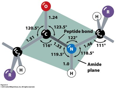
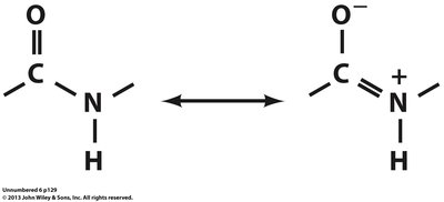
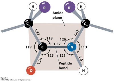
The peptide bond exhibits partial double-bond character due to resonance, resulting in a planar structure and restricted rotation. This planarity is crucial for the formation of regular secondary structures.
Planar Structure: Six atoms (Cα, C, O, N, H, Cα) lie in the same plane.
Restricted Rotation: Rotation is limited around the peptide bond, but allowed around the bonds flanking the Cα atom (φ and ψ angles).
Resonance: The peptide bond is stabilized by resonance between the carbonyl and amide groups.
Rotation Around the Peptide Backbone
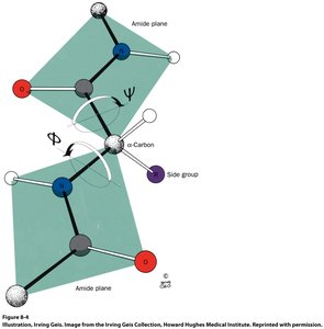
Rotation in the polypeptide backbone occurs around the bonds adjacent to the Cα atom, defined by the dihedral angles φ (phi) and ψ (psi). These angles determine the conformational flexibility and are critical for secondary structure formation.
Dihedral Angles: φ (N–Cα) and ψ (Cα–C) describe the rotation around the backbone.
Conformational Constraints: Steric hindrance and the nature of the side chain (R group) limit the allowed values of φ and ψ.
Main Chain and Side Chain Organization
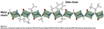
The polypeptide backbone consists of repeating units, with side chains (R groups) projecting from the main chain. The arrangement of these groups influences protein folding and function.
Main Chain: The repeating N–Cα–C units form the backbone.
Side Chains: R groups are attached to the Cα atom and vary among amino acids.
Summary Table: Secondary Structure Dihedral Angles
Different secondary structures are characterized by specific φ and ψ angles. The table below summarizes these values for common protein secondary structures.
Secondary Structure | φ (deg) | ψ (deg) |
|---|---|---|
Right-handed α helix (α) | -57 | -47 |
Parallel β pleated sheet (↑↑) | -119 | 113 |
Antiparallel β pleated sheet (↑↓) | -139 | 135 |
Right-handed 310 helix (3) | -49 | -26 |
Right-handed π helix (π) | -57 | -70 |
2.21 ribbon (2) | -78 | 59 |
Left-handed polyglycine II and polyproline II helices (II) | -79 | 150 |
Collagen (C) | -51 | 153 |
Left-handed α helix (αL) | 57 | 47 |
Ramachandran Plot
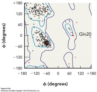
The Ramachandran plot visualizes the allowed regions of φ and ψ angles for amino acid residues in proteins. It is a fundamental tool for understanding protein structure and predicting secondary structure elements.
Allowed Regions: Most residues fall within energetically favorable regions corresponding to α-helices and β-sheets.
Glycine and Proline: These amino acids have unique conformational properties, often found outside typical regions.
Summary
Understanding the hierarchical structure of proteins and the chemistry of peptide bonds is essential for grasping how proteins fold and function. The interplay between primary sequence, peptide bond properties, and backbone dihedral angles underlies the formation of complex protein structures.
