 Back
BackSensory Transduction: Mechanisms and Molecular Basis
Study Guide - Smart Notes
 Tailored notes based on your materials, expanded with key definitions, examples, and context.
Sensory Transduction
Introduction to Sensory Transduction
Sensory transduction is the process by which environmental stimuli are converted into electrical signals by specialized cells. This fundamental process underlies all sensory perception, including vision, hearing, touch, taste, and smell, as well as other modalities such as pain, temperature, and proprioception.
Stimulus Detection: Environmental stimuli (e.g., photons, pressure, chemicals) are detected by receptor molecules in sensory cells.
Signal Conversion: Receptor activation leads to the opening or closing of transduction channels, altering the cell's membrane potential.
Neural Response: Changes in membrane potential modulate the firing rate or neurotransmitter release, transmitting information to the nervous system.
Examples of Sensory Transducing Cells:
Hair cells of the ear (mechanoreceptors)
Rods and cones of the retina (photoreceptors)
Taste receptors of the tongue (chemoreceptors)
Odor receptors of the nose (chemoreceptors)
Somatosensory receptors in the skin (mechanoreceptors)
Thermoceptors, nociceptors, and multimodal receptors
Phototransduction
Mechanism of Phototransduction in Vertebrates
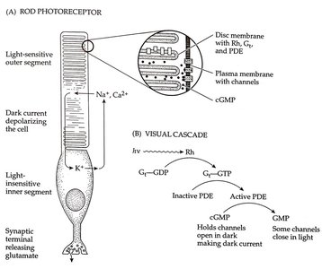
Phototransduction is the process by which light is converted into electrical signals in the retina. In vertebrates, this process occurs in rod and cone photoreceptors and relies on G-protein coupled receptors (GPCRs) called rhodopsins.
Light Detection: Photons activate rhodopsin (Rh), a GPCR located in the outer segment of rods and cones.
Signal Cascade: Activated rhodopsin stimulates the G-protein transducin (Gt), which in turn activates phosphodiesterase (PDE).
cGMP Breakdown: PDE hydrolyzes cGMP, reducing its concentration.
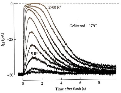
Channel Closure: Lower cGMP levels cause cyclic nucleotide-gated (CNG) channels to close, reducing Na+ and Ca2+ influx.
Hyperpolarization: The cell hyperpolarizes, leading to closure of voltage-gated Ca2+ channels and decreased glutamate release.
Key Equations:
Activation:
G-protein cycle:
PDE activation:
cGMP hydrolysis:
Functional Consequences:
In darkness, CNG channels are open, and the cell is depolarized, continuously releasing glutamate.
Light exposure closes CNG channels, hyperpolarizing the cell and reducing glutamate release.
Alternative Phototransduction Mechanisms
Other organisms utilize different molecular strategies for phototransduction:
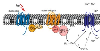
Drosophila: Use a Gq-coupled receptor that activates phospholipase C (PLC), leading to the opening of TRP channels and cell depolarization.
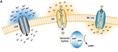
Prokaryotes: Employ light-gated ion channels or pumps such as channelrhodopsin (ChR), halorhodopsin (HR), and bacteriorhodopsin (BR).
Taste Transduction
Mechanisms of Taste Transduction
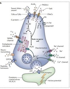
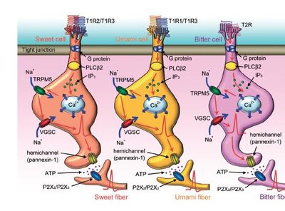
Taste buds contain specialized receptor cells that convert chemical tastants into electrical signals. Different types of taste cells detect sweet, sour, salty, bitter, and umami stimuli via distinct molecular mechanisms.
Type II Taste Cells: Detect sweet, umami, and bitter via GPCRs (T1Rs and T2Rs) that activate Gq proteins, leading to IP3-mediated Ca2+ release and opening of TRPM5 channels, resulting in depolarization and ATP release.
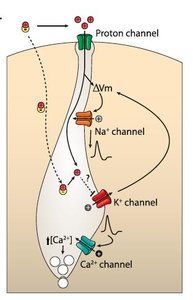
Type III Taste Cells: Detect sour and salty via direct ion channel activation (e.g., Otop1 for H+, ENaC for Na+), leading to depolarization, action potential firing, and neurotransmitter release.
Otop1: The H+-Permeable Channel in Sour Taste
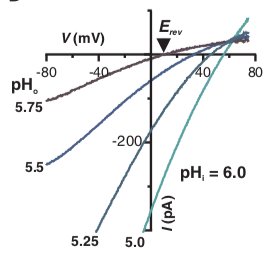
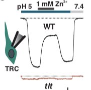
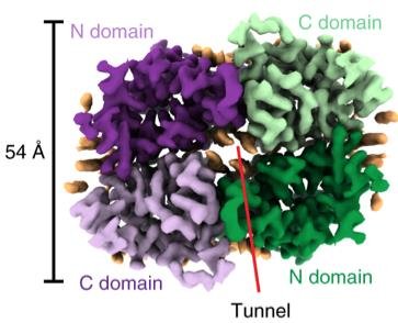
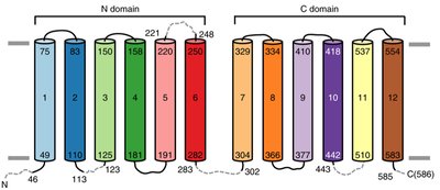
Otop1 is a proton-selective ion channel essential for sour taste perception. Identified through genetic screening, Otop1 is sensitive to Zn2+ and is required for acid detection in taste cells.


Somatosensation and Mechanotransduction
Somatosensory Receptors
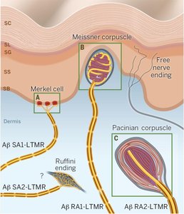
Somatosensory receptors are specialized mechanoreceptors that detect touch, pressure, vibration, and stretch. Examples include Meissner corpuscles (light touch), Merkel cells (texture), Pacinian corpuscles (vibration), and Ruffini endings (stretch).
Merkel Cells and Piezo2 Channels
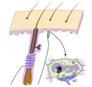
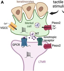
Merkel cells are mechanoreceptors densely located in areas such as the fingertips and lips. They express Piezo2, a pressure-activated cation channel essential for detecting light touch and texture.
Mechanism: Pressure opens Piezo2 channels, depolarizing the Merkel cell and leading to Ca2+ influx and neuropeptide release. This excites adjacent sensory nerve fibers.
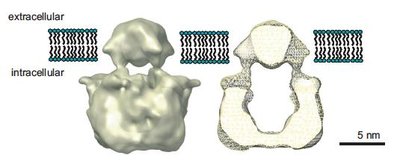
Piezo2 Structure: Piezo proteins are large, with three subunits forming a trimeric channel complex.
Thermosensation and TRP Channels
TRP Channels as Temperature Sensors
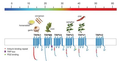
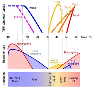
Transient Receptor Potential (TRP) channels are a family of cation channels that transduce temperature and chemical stimuli. Different TRP channels are activated by specific temperature ranges and chemical ligands.
Cold Sensors: TRPM8 and TRPA1
Heat Sensors: TRPV1, TRPV2, TRPV3
Channel Properties: Most TRP channels are non-selective cation channels, permeable to Na+ and Ca2+.
TRP Channel Structure and Diversity
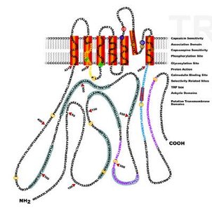
TRP channels share structural similarities with voltage-gated ion channels, including six transmembrane segments per subunit and a tetrameric assembly. They are gated by temperature, ligands, and other factors.
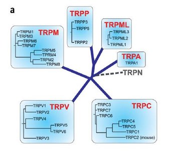
Subfamilies: TRPC (canonical), TRPM (melastatin), TRPV (vanilloid), TRPA (ankyrin), TRPP, TRPML
Functional Diversity: TRP channels are involved in sensory transduction, osmoregulation, and other cellular processes.
Summary Table: Sensory Transduction Mechanisms
Modality | Receptor Type | Transduction Mechanism | Key Ion Channels | Effect |
|---|---|---|---|---|
Vision (Vertebrate) | Photoreceptor (GPCR) | cGMP-mediated channel closure | CNG, Cav1.4 | Hyperpolarization |
Vision (Invertebrate) | GPCR/TRP | PLC/IP3 pathway | TRP | Depolarization |
Taste (Type II) | GPCR | IP3-mediated Ca2+ release | TRPM5, Nav | Depolarization |
Taste (Type III) | Ion channel | Direct ion influx/block | Otop1, ENaC, Nav | Depolarization |
Touch | Mechanoreceptor | Pressure-gated channel | Piezo2 | Depolarization |
Temperature | TRP channel | Temperature/ligand gating | TRPV, TRPM, TRPA | Depolarization |
