 Back
Structural Biology Techniques in Protein Biochemistry
Study Guide - Smart Notes
 Tailored notes based on your materials, expanded with key definitions, examples, and context.
Structural Biology: Experimental Approaches to Protein Structure
Sample Requirements for Structure Determination
Determining the three-dimensional structure of proteins requires careful sample preparation. The requirements differ among major techniques:
X-ray Crystallography: Requires highly pure, concentrated protein (typically 5–20 mg/mL) and a known sequence.
NMR Spectroscopy: Requires pure protein, typically 0.5–1 mM (10–20 mg/mL for a 20 kDa protein), and isotopic labeling with 15N and 13C.
Cryo-EM: Requires much less protein, often well below 1 mg/mL, and can tolerate lower purity.
Example: For membrane proteins, cryo-EM is often preferred due to difficulties in crystallization.
Circular Dichroism (CD) Spectroscopy and Secondary Structure Analysis
Principle and Applications
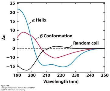
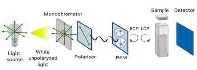
CD spectroscopy is a rapid method for analyzing protein secondary structure. It exploits the chirality of proteins and the arrangement of peptide bonds in α-helices, β-sheets, and random coils. CD measures the difference in absorption of right and left circularly polarized light as a function of wavelength, primarily in the far-UV (190–250 nm).
α-Helix: Characteristic negative bands near 208 and 222 nm.
β-Sheet: Single negative band near 218 nm.
Random Coil: Strongly negative band near 195–200 nm.
Mutagenesis: Used to test if amino acid changes disrupt secondary structure.
Thermal Stability: CD can monitor protein unfolding by measuring changes in signal at specific wavelengths during heating.
Example: CD spectra distinguish between α-helical, β-sheet, and random coil proteins.
Dynamic Light Scattering (DLS) and Quaternary Structure Detection
Principle and Applications
DLS is used to measure the hydrodynamic radius of proteins in solution, providing information about quaternary structure and aggregation. A laser is shone through the sample, and fluctuations in scattered light intensity (due to Brownian motion) are analyzed to determine the diffusion coefficient, which is converted to radius using the Stokes–Einstein equation:
Monodisperse vs. Polydisperse: DLS distinguishes homogeneous samples from those containing aggregates.
Resolving Power: Can detect large aggregates but cannot reliably distinguish monomers from dimers.
Clinical Application: Used in quality control for biologic drugs to detect aggregation.
Example: DLS and CD are paired to confirm protein structure and aggregation status in drug formulations.
Chemical Crosslinking for Quaternary Structure
Principle and Applications
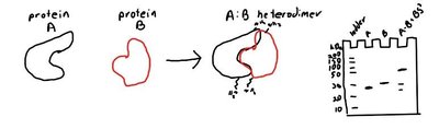
Chemical crosslinking uses bifunctional reagents (e.g., BS3) that react with primary amines (lysine residues) to form stable amide bonds, locking protein complexes in place. SDS-PAGE analysis then reveals the presence of higher-order structures.
Crosslinker: BS3 contains two NHS-ester groups reactive with amines.
Application: Used to confirm oligomeric states and protein–protein interactions.
Cryoelectron Microscopy (Cryo-EM)
Principle and Applications
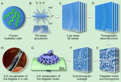
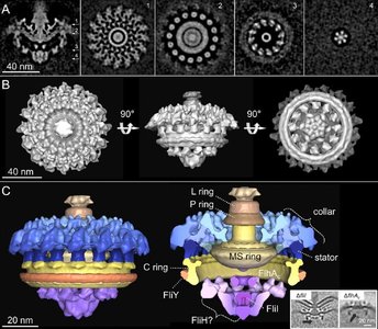
Cryo-EM is a transmission electron microscopy technique where samples are rapidly frozen in vitreous ice, preserving native structure. Two main approaches:
Single-Particle Analysis: Images thousands of particles in random orientations for 3D reconstruction.
Cryo-Electron Tomography: Tilts the sample to collect a series of images for 3D reconstruction of structures within cells.
Recent advances allow near-atomic resolution (1.2–3 Å). Cryo-EM is especially suited for large, flexible, glycosylated proteins.
Example: Cryo-EM was used to determine the structure of the SARS-CoV-2 spike protein, guiding vaccine design.
X-Ray Crystallography
Principle and Workflow
X-ray crystallography is the most established method for high-resolution protein structure determination. The workflow includes:
Protein Purification: Requires highly pure, concentrated protein.
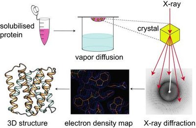
Crystallization: Often uses vapor diffusion to grow crystals.
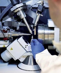
X-ray Diffraction: Crystals are exposed to X-rays, and diffraction patterns are collected.
Electron Density Map: Intensities and phases are used to reconstruct the map; the phase problem is solved by molecular replacement or experimental methods.
Model Building: Amino acid sequence is threaded into the density map and refined.
Advantages: Atomic resolution, well-established computational tools.
Limitations: Membrane proteins are difficult to crystallize; disordered regions are invisible; crystal form may differ from solution form.
Resolution and R-factor
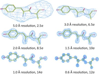
Resolution (in Å or nm) indicates the level of detail visible in the electron density map. Atomic resolution is typically achieved at 1.2 Å or better. The R-factor measures agreement between observed and calculated diffraction data; lower values indicate better models.
Nuclear Magnetic Resonance (NMR) Spectroscopy
Principle and Applications
NMR spectroscopy determines protein structure in solution, providing information on dynamics and disordered regions. Requires isotopic labeling with 15N and 13C for larger proteins. Multi-dimensional experiments are essential for structure determination.
Advantages: No need for crystallization; can observe dynamics and disordered regions.
Limitations: Not suitable for insoluble proteins; size limit (~45 kDa).
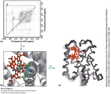
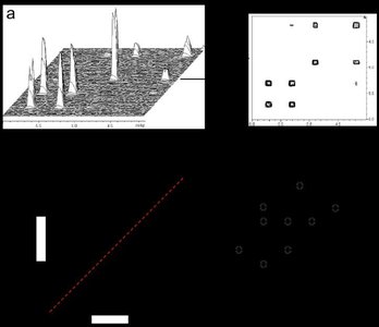
COSY: Correlation Spectroscopy
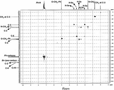
COSY detects through-bond correlations between protons up to three bonds apart. Cross-peaks off the diagonal indicate coupled protons, aiding in peak assignment and structure determination.
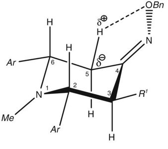
NOESY: Nuclear Overhauser Effect Spectroscopy
NOESY detects through-space correlations between protons within ~5 Å. Cross-peak intensity falls off with the sixth power of distance (), providing distance restraints for 3D structure calculation.
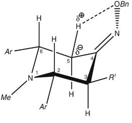
HSQC: Heteronuclear Single Quantum Coherence Spectroscopy
HSQC detects correlations between different nuclei (e.g., 1H–13C or 1H–15N). The 1H–15N HSQC spectrum serves as a protein fingerprint, with each cross-peak representing an amide N–H. Peak shifts upon ligand binding map the binding site.
Combining Cryo-EM and X-ray Crystallography
Principle and Applications
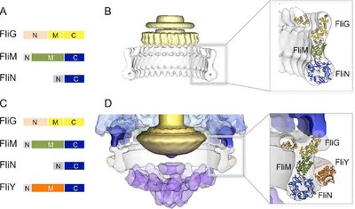
Combining cryo-EM (for large complexes) with high-resolution crystal or NMR structures (for individual proteins) enables visualization of multi-protein assemblies at near-atomic detail. Crystal structures are fit into lower-resolution cryo-EM maps.
AlphaFold: AI-Based Structure Prediction
Principle and Impact
AlphaFold is an AI tool that predicts protein structures from amino acid sequences with high accuracy. AlphaFold2 solved the long-standing problem of reliable structure prediction, and AlphaFold3 extends this to protein complexes and interactions with nucleic acids and small molecules. Limitations remain for post-translational modifications and large assemblies, but the tool has revolutionized structural biology.
Applications: Used for structure prediction, guiding experiments, and drug design.
Limitations: Some modifications and alternative conformations are not fully captured.
Example: AlphaFold models are now used for molecular replacement in crystallography.
Summary Table: Comparison of Structural Biology Techniques
Technique | Sample Requirement | Resolution | Strengths | Limitations |
|---|---|---|---|---|
X-ray Crystallography | High purity, concentrated, known sequence | Atomic (1.2 Å or better) | High resolution, established methods | Crystallization required, disordered regions invisible |
NMR Spectroscopy | High purity, isotopic labeling, soluble | Atomic for small proteins | Solution structure, dynamics, disordered regions visible | Size limit (~45 kDa), insoluble proteins inaccessible |
Cryo-EM | Lower concentration, less purity | Near-atomic (1.2–3 Å) | Large complexes, membrane proteins, no crystallization | Lower resolution for flexible complexes |
CD Spectroscopy | Chiral, absorbs in UV | Secondary structure only | Rapid, no sequence needed | No atomic detail |
DLS | Clean sample | Hydrodynamic radius | Detects aggregation, size | Limited resolving power |
Chemical Crosslinking | Reactive groups | Quaternary structure | Confirms oligomeric state | Indirect, requires follow-up analysis |
