 Back
BackChromosomes, Mitosis, and Meiosis: Structure and Function of Genetic Material
Study Guide - Smart Notes
 Tailored notes based on your materials, expanded with key definitions, examples, and context.
Chromosomes, Mitosis, and Meiosis
Historical Perspective
Understanding the molecular basis of heredity began in the early 20th century, when scientists established that genes are located on chromosomes within the nucleus and that chromosomes are composed of DNA. The discovery of the double-helix structure of DNA by James Watson and Francis Crick in 1953, based on Rosalind Franklin's work, revolutionized our understanding of genetic information storage and transmission.
Structure of Nucleic Acids
Nucleotides: The Building Blocks
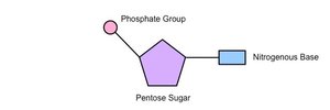
Nucleic acids, including DNA and RNA, are polymers made up of repeating units called nucleotides. Each nucleotide consists of three components:
Pentose sugar (deoxyribose in DNA, ribose in RNA)
Phosphate group
Nitrogenous base
Genetic material exists in the form of nucleic acids, which are essential for storing and transmitting hereditary information.
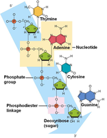
Nitrogenous Bases
There are two types of nitrogenous bases in nucleic acids:
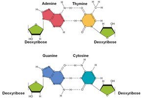
Purines: Double-ring structures (Adenine [A], Guanine [G])
Pyrimidines: Single-ring structures (Cytosine [C], Thymine [T], Uracil [U])
DNA contains A, T, C, and G; RNA contains A, U, C, and G.
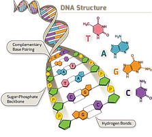
DNA Structure
DNA is a double helix, resembling a spiral staircase. Each strand is a polymer of nucleotides linked by phosphodiester bonds. The backbone consists of alternating phosphate and deoxyribose sugar units, while the steps are pairs of nitrogenous bases.
Base pairing is specific: A pairs with T (via two hydrogen bonds), and G pairs with C (via three hydrogen bonds). This complementary base pairing allows for accurate DNA replication.
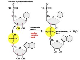
Phosphodiester Bonds
Nucleotides are joined by phosphodiester bonds between the 3' hydroxyl group of one sugar and the 5' phosphate group of the next. This linkage forms the sugar-phosphate backbone of DNA and RNA.
DNA Base Pairing and Chargaff's Rules
In double-stranded DNA, the number of purines equals the number of pyrimidines (A = T, G = C). This is known as Chargaff's rules and is essential for the double helix's stability and replication fidelity.
DNA Organization in Cells
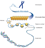
Chromatin, Chromosomes, and Genes
In eukaryotic cells, DNA is packaged with proteins (histones) into a complex called chromatin. During cell division, chromatin condenses to form visible chromosomes. Each chromosome contains many genes, which are sequences of DNA that encode proteins or functional RNA molecules.
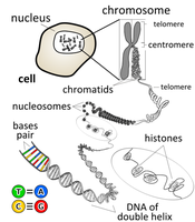
Nucleosomes and Chromosome Structure
DNA wraps around histone proteins to form nucleosomes, which further coil and fold to produce the highly compact structure of chromosomes. This organization allows efficient packaging and regulation of genetic material.
The Cell Cycle
Phases of the Cell Cycle
The cell cycle is the sequence of events from one cell division to the next. It consists of:
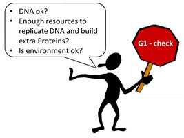
Interphase: G1 (growth), S (DNA synthesis), G2 (preparation for division)
Mitotic (M) phase: Mitosis (nuclear division) and cytokinesis (cytoplasmic division)
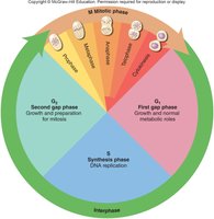
Checkpoints (e.g., G1 and G2) ensure the cell is ready to proceed to the next phase, maintaining genomic integrity.
Mitosis
Overview and Functions
Mitosis is the process by which a eukaryotic cell divides its nucleus, producing two genetically identical daughter cells. It is essential for growth, tissue repair, and asexual reproduction in multicellular organisms.
Phases of Mitosis
Prophase: Chromatin condenses into chromosomes; nuclear envelope breaks down; spindle fibers form.

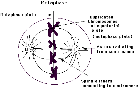
Metaphase: Chromosomes align at the cell's equator; spindle fibers attach to kinetochores.
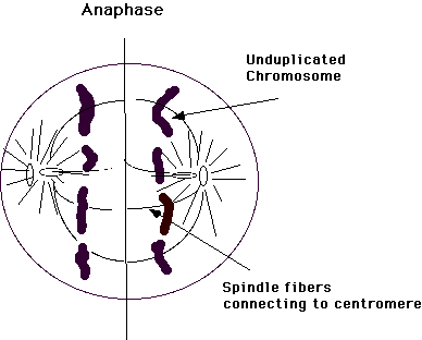
Anaphase: Sister chromatids separate and move toward opposite poles.
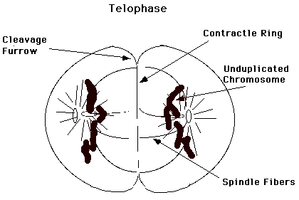
Telophase: Chromosomes decondense; nuclear envelopes reform; spindle disappears.
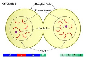
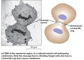
Cytokinesis
Cytokinesis is the division of the cytoplasm, resulting in two separate daughter cells. In animal cells, a cleavage furrow forms, while in plant cells, a cell plate develops.
Prokaryotic Cell Division
Binary Fission
Prokaryotes (bacteria and archaea) divide by binary fission, a simpler process than mitosis. The DNA replicates, and the cell splits into two genetically identical daughter cells. This process is rapid and efficient, allowing for quick population growth.
Chromosome Number and Ploidy
Homologous Chromosomes, Diploid and Haploid Cells
Somatic cells contain pairs of homologous chromosomes (one from each parent). The diploid (2n) number refers to cells with two sets of chromosomes, while haploid (n) cells (e.g., gametes) have one set. Fusion of gametes during fertilization restores the diploid number in the zygote.
Polyploidy
Polyploidy refers to cells with three or more sets of chromosomes. It is common in plants and can result in increased size and vigor.
Meiosis
Overview and Significance
Meiosis is a specialized cell division that reduces the chromosome number by half, producing four genetically unique haploid cells (gametes). It consists of two sequential divisions: Meiosis I and Meiosis II. Meiosis introduces genetic variation through independent assortment and crossing-over.
Stages of Meiosis
Meiosis I: Homologous chromosomes pair, undergo crossing-over, and then separate into different cells.
Meiosis II: Sister chromatids separate, similar to mitosis, resulting in four haploid cells.
Genetic Variation
Crossing-over during Prophase I and the random assortment of chromosomes during Metaphase I generate genetic diversity among gametes.
Sexual Life Cycles
Animals, Plants, and Fungi
Sexual life cycles vary among eukaryotes:
Animals: Diploid somatic cells; only gametes are haploid.
Plants: Alternation of generations between multicellular diploid (sporophyte) and haploid (gametophyte) stages.
Fungi and Protists: Often have dominant haploid stages.
These cycles ensure genetic diversity and adaptation to changing environments.
Key Terms and Definitions
Chromosome: A DNA molecule with associated proteins, carrying genetic information.
Chromatin: The complex of DNA and proteins forming chromosomes.
Chromatid: One of two identical halves of a replicated chromosome.
Sister chromatids: Two identical chromatids joined at a centromere.
Nucleosome: DNA wrapped around histone proteins.
Histones: Proteins that package and order DNA into nucleosomes.
Gene: A segment of DNA encoding a functional product.
Kinetochore: Protein structure on the centromere where spindle fibers attach.
Centromere: Region where sister chromatids are joined.
