 Back
BackInside the Cell: Structure and Function of Cellular Components
Study Guide - Smart Notes
 Tailored notes based on your materials, expanded with key definitions, examples, and context.
Inside the Cell
Introduction
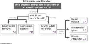
This chapter explores the internal structure of cells, emphasizing how the collaboration of cellular components underlies the properties of life. It compares prokaryotic and eukaryotic cells, examines the organization and function of organelles, and details the dynamic systems that maintain cellular function.
Prokaryotic Cell Structures and Their Functions
Overview of Prokaryotic Cells
Prokaryotic cells, which include Bacteria and Archaea, lack a membrane-bound nucleus but possess a variety of specialized structures:
Chromosome: Usually a single, circular DNA molecule associated with proteins, located in a region called the nucleoid.
Plasmids: Small, circular DNA molecules that carry genes beneficial for survival.
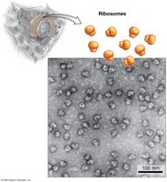
Ribosomes: Macromolecular complexes of RNA and protein that synthesize proteins.
Cytoplasm: The internal fluid containing all cell contents.
Cell Wall: Provides structural support and shape; in bacteria, primarily composed of peptidoglycan.
Plasma Membrane: Selectively permeable barrier composed of phospholipids (fatty acids in Bacteria, branched isoprenoids in Archaea).
Photosynthetic Membranes: In some species, internal membranes for photosynthesis.
Organelles: Some bacteria have membrane-bound compartments for specialized functions (e.g., storing ions, orienting with magnetite crystals).
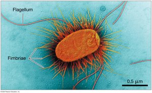
External Structures: Flagella (movement) and fimbriae (attachment).
Eukaryotic Cell Structures and Their Functions
General Features of Eukaryotic Cells
Eukaryotic cells are generally larger and more complex than prokaryotic cells. They contain numerous membrane-bound organelles that compartmentalize cellular functions:
Nucleus: Stores genetic information and is the site of RNA synthesis and ribosome assembly.
Ribosomes: Sites of protein synthesis, found free in the cytosol or bound to the endoplasmic reticulum (ER).
Endoplasmic Reticulum (ER): Rough ER (with ribosomes) synthesizes and processes proteins; Smooth ER (without ribosomes) synthesizes lipids and detoxifies molecules.
Golgi Apparatus: Processes, sorts, and ships proteins and lipids received from the ER.
Lysosomes: Contain hydrolytic enzymes for digestion and recycling (mainly in animal cells).
Vacuoles: Storage organelles in plants, fungi, and some protists; roles include storage of water, ions, pigments, and defensive compounds.
Peroxisomes: Centers for oxidation-reduction reactions; detoxify harmful substances.
Mitochondria: Sites of ATP production via cellular respiration; contain their own DNA and ribosomes.
Chloroplasts: Sites of photosynthesis in plants and algae; contain their own DNA and ribosomes.
Cytoskeleton: Network of protein fibers that provides structural support, organizes organelles, and facilitates movement.
Cell Wall: Found in plants, fungi, and algae; provides structural support and protection.
Cellular Compartmentalization and Function
Benefits of Organelles
Organelles allow eukaryotic cells to compartmentalize incompatible chemical reactions and increase the efficiency of cellular processes. The cytosol, the fluid portion of the cell, occupies a small volume, which helps offset the limitations of surface-area-to-volume ratio in larger cells.
Cell Systems: Integration of Cellular Components
Nuclear Transport
The nucleus is separated from the cytoplasm by the nuclear envelope, which contains nuclear pore complexes. These complexes regulate the selective import and export of molecules. Proteins destined for the nucleus contain a nuclear localization signal (NLS), a specific amino acid sequence that acts as a molecular "zip code" for nuclear import.
The Endomembrane System
The endomembrane system includes the ER, Golgi apparatus, lysosomes, and associated vesicles. It is responsible for the synthesis, processing, sorting, and transport of proteins and lipids. Proteins destined for secretion or specific organelles are synthesized with signal sequences that direct them to the correct location.
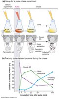
PULSE-CHASE EXPERIMENT: Used to track the movement of proteins through the endomembrane system. Proteins are labeled during a short "pulse" and their movement is followed during the "chase." Results show proteins move from the rough ER to the Golgi apparatus and then to their final destinations.
Protein Targeting and Sorting
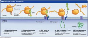
The signal hypothesis explains how proteins are directed to the ER. A 20-amino-acid ER signal sequence is recognized by a signal recognition particle (SRP), which directs the ribosome to the ER membrane. The protein is then translocated into the ER lumen, where it is folded and modified (e.g., glycosylation).
Vesicular Transport and Exocytosis
Proteins are transported between organelles in membrane-bound vesicles. Specific molecular tags ensure that vesicles deliver their cargo to the correct destination. Exocytosis is the process by which vesicles fuse with the plasma membrane to release their contents outside the cell.
Lysosomal Recycling Pathways
Lysosomes recycle cellular materials via three main pathways:
Receptor-mediated endocytosis: Uptake of specific molecules via receptor binding and vesicle formation.
Phagocytosis: Engulfment of large particles or cells to form a phagosome, which fuses with a lysosome for digestion.
Autophagy: Degradation of the cell's own damaged organelles or macromolecules within autophagosomes that fuse with lysosomes.
The Dynamic Cytoskeleton
Types of Cytoskeletal Elements
The cytoskeleton is composed of three main types of protein filaments:
Actin Filaments (Microfilaments): Smallest filaments, composed of actin; involved in cell shape, movement, and division.
Intermediate Filaments: Provide mechanical strength; include keratins and nuclear lamins.
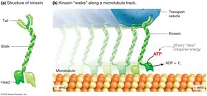
Microtubules: Largest filaments, composed of tubulin dimers; serve as tracks for vesicle transport and are essential for chromosome separation during cell division.
Microtubule-Based Transport
Microtubules serve as tracks for the movement of vesicles and organelles. The motor protein kinesin "walks" along microtubules, converting ATP energy into mechanical work to transport cargo.
Flagella and Cilia
Flagella and cilia are cellular appendages used for movement. Prokaryotic flagella rotate, while eukaryotic flagella and cilia have a "9+2" arrangement of microtubules and move by bending, powered by the motor protein dynein.
Summary Table: Eukaryotic Cell Components
Organelle | Main Function | Key Features |
|---|---|---|
Nucleus | Information storage, ribosome assembly | Double membrane, nuclear pores, nucleolus |
Ribosomes | Protein synthesis | Free or bound to ER, not membrane-bound |
Rough ER | Protein synthesis and processing | Studded with ribosomes |
Smooth ER | Lipid synthesis, detoxification | Lacks ribosomes |
Golgi Apparatus | Protein and lipid modification, sorting, shipping | Stacked cisternae, cis/trans polarity |
Lysosomes | Digestion and recycling | Acidic interior, hydrolytic enzymes |
Vacuoles | Storage, digestion, defense | Large in plant cells |
Peroxisomes | Redox reactions, detoxification | Contain catalase |
Mitochondria | ATP production | Double membrane, own DNA |
Chloroplasts | Photosynthesis | Three membranes, own DNA |
Cytoskeleton | Structural support, movement | Actin, intermediate filaments, microtubules |
Cell Wall | Protection, support (plants, fungi, algae) | Cellulose, chitin, or other polysaccharides |
Conclusion
The structure and function of cellular components are intricately linked, allowing cells to perform the complex processes necessary for life. Understanding these systems provides a foundation for further study in cell biology, physiology, and molecular biology.
