 Back
BackL 5.1: Protozoan Protists: Diversity, Structure, and Function
Study Guide - Smart Notes
 Tailored notes based on your materials, expanded with key definitions, examples, and context.
L 5.1
Protozoan Protists: Introduction and Overview
Protozoan protists are a diverse group of single-celled eukaryotic organisms, historically classified as 'animal-like' protists due to their heterotrophic lifestyle. They are found in a wide range of environments and play important ecological and medical roles. The study of protozoan protists has been foundational in cell biology and evolutionary studies.
Size: Typically 10–50 micrometers.
Discovery: First observed by Anton van Leeuwenhoek in 1674.
Classification: The term 'protozoa' was coined by Georg Goldfuss in 1818; early microscopists referred to them as 'animalcules.'
Major Groups: Amoebozoa, Excavata, Rhizaria, and Alveolata.
Additional info: Protozoan protists are crucial for understanding eukaryotic evolution and the origins of multicellularity.
Phylum Amoebozoa
General Characteristics
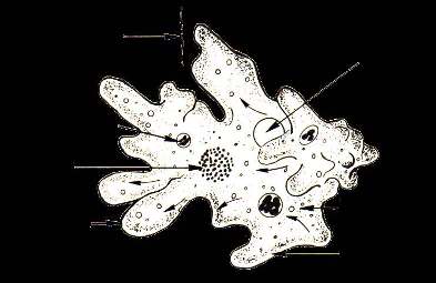
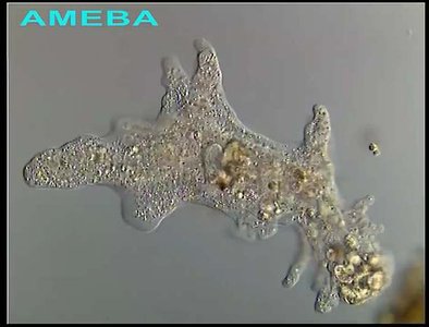
Amoebozoans are characterized by their lobe- or tube-shaped pseudopodia (lobopodia), which are used for movement and feeding. Most are unicellular, though some form multicellular or multinucleate structures (slime molds).
Locomotion: Achieved via sol-gel conversion, involving cytoplasmic streaming regulated by osmotic pressure and ionic changes.
Mitochondria: Most have branching tubular cristae; some, like Entamoeba, have lost mitochondria.
Flagella: Generally absent except in some slime molds and Entamoeba gametes.
Class Tubulinea
Subclass Gymnamoebia (Lobosa): Includes common freshwater amoebae and testate amoebae.
Genus Amoeba: Free-living, found in freshwater environments.
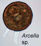
Genus Arcella: Testate amoebae with a circular test and finger-like pseudopods.
Genus Difflugia
These amoebae produce shells (tests) from sand granules, which are passed to daughter cells during division. Common in marshes.
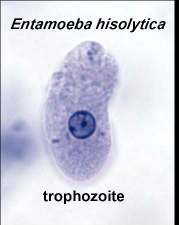
Genus Entamoeba
Amitochondriate: Lacks mitochondria.
Parasitic: Includes important human parasites.
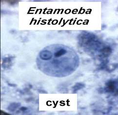
Entamoeba histolytica: Causes amoebic dysentery; infects ~50 million people worldwide.
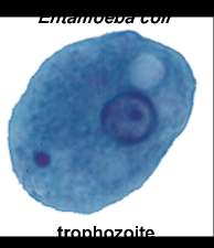
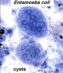
Entamoeba coli: Non-pathogenic, but important for differential diagnosis in stool samples.
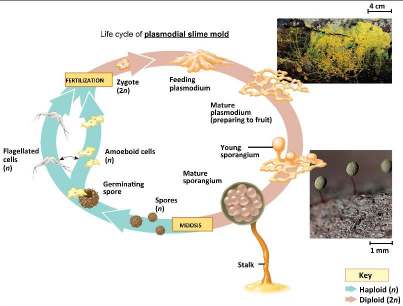
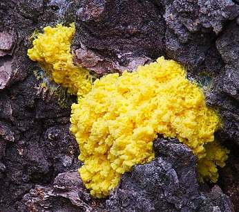
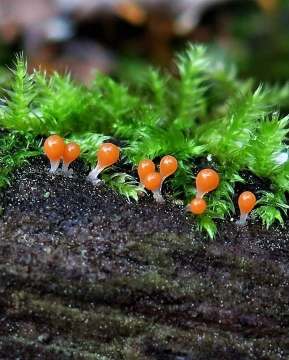
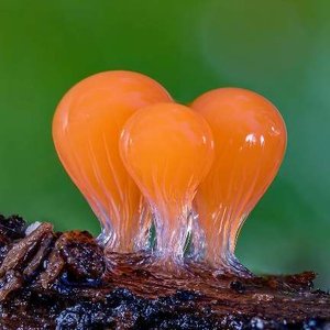
Slime Molds (Infra Phylum Mycetozoa)
Slime molds were once classified as fungi but are now placed in Amoebozoa based on molecular data. They are divided into plasmodial and cellular slime molds.
Plasmodial Slime Molds: Multinucleate, brightly pigmented (yellow/orange), form a large mass called a plasmodium.
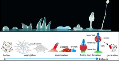
Cellular Slime Molds: Aggregate into multicellular structures; model organism: Dictyostelium discoideum.
Kingdom Excavata: Flagellates
Phylum Metamonada
Class Parabasalia: Amitochondriate, anaerobic flagellates, often symbionts or parasites.
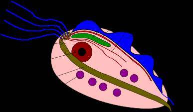
Trichomonas sp.: Human pathogen; features include anterior flagella, undulating membrane, parabasal body, and hydrogenosomes (ATP production).
Order Diplomonadida
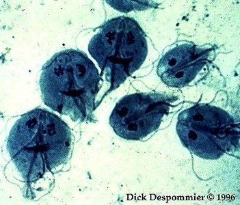
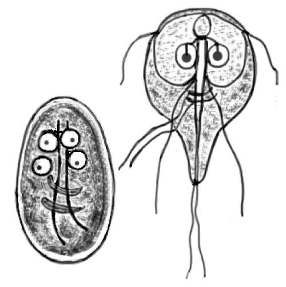
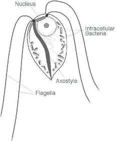
Giardia intestinalis (G. lamblia): Common intestinal parasite; lacks mitochondria and Golgi, has mitosomes, and two nuclei.
Order Oxymonadida
Flagellated protozoa found in termite intestines, aiding in cellulose digestion via symbiotic bacteria.
Phylum Euglenozoa
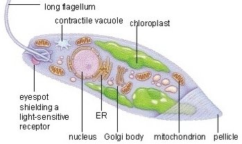
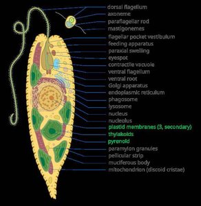
With chloroplasts: Euglena (mixotrophic, photosynthetic and heterotrophic).
Without chloroplasts: Trypanosoma (Chagas disease, sleeping sickness), Leishmania (leishmaniasis).
Flagella: Unique structure with paraflagellar rod next to 9+2 microtubule arrangement.
Class Euglenoidea
Mixotrophy: Capable of both phagocytosis and photosynthesis (chlorophyll a and b, carotenoids).
Eyespot: Light-sensitive organelle for phototaxis.
Pellicle: Protein bands beneath the plasma membrane, providing flexibility and shape.
Paramylon granules: Storage polysaccharide (β 1,3-linked glucan).
Class Kinetoplastea
Kinetoplast: DNA-containing granule within a single mitochondrion, unique to this group.
Trypanosoma: Causes Chagas disease and sleeping sickness.
Leishmania: Causes leishmaniasis.
Rhizaria
Rhizarians are amoeboid protists with fine, reticulating pseudopods and often elaborate mineral skeletons (tests).
Foraminifera (Forams): Marine, produce multi-chambered tests of calcium carbonate; important in paleontology.
Radiolaria: Marine plankton with intricate silica skeletons; their remains form radiolarian ooze on the ocean floor.
Alveolata
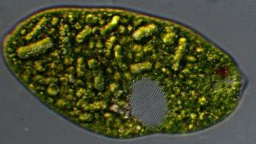
Phylum Ciliophora (Ciliates)
Cilia: Short, hair-like organelles for movement and feeding; more numerous and shorter than flagella.
Examples: Paramecium, Spirostomum, Colpidium, Vorticella, Stentor, Euplotes, Balantidium coli.
Kleptoplasts: Some ciliates retain functional plastids from ingested algae.
Phylum Apicomplexa (Sporozoans)
Apicoplast: Unique organelle involved in host cell penetration; nonphotosynthetic plastid.
Parasitic: All are animal parasites; important genera include Plasmodium (malaria), Toxoplasma (toxoplasmosis), and Cryptosporidium (cryptosporidiosis).
Malaria Life Cycle: Involves two hosts (mosquito and human); complex cycle with liver and blood stages.
Malaria Life Cycle Key Steps:
Infected mosquito injects sporozoites into human.
Sporozoites infect liver cells, mature into schizonts, and release merozoites.
Merozoites infect red blood cells, multiply asexually, and cause symptoms.
Some merozoites develop into gametocytes, which are taken up by mosquitoes, continuing the cycle.
Additional info: Plasmodium vivax and P. ovale can form dormant liver stages (hypnozoites), causing relapses.
Summary Table: Key Protozoan Protist Groups
Group | Main Features | Examples | Medical/Ecological Importance |
|---|---|---|---|
Amoebozoa | Lobopodia, sol-gel movement, some form tests | Amoeba, Arcella, Entamoeba, slime molds | Pathogens (E. histolytica), model for multicellularity (Dictyostelium) |
Excavata | Flagellates, some lack mitochondria, unique organelles | Trichomonas, Giardia, Euglena, Trypanosoma | Human pathogens (Giardia, Trypanosoma) |
Rhizaria | Reticulating pseudopods, mineral skeletons | Foraminifera, Radiolaria | Marine ecology, paleontology |
Alveolata | Cilia or apical complex, alveoli under membrane | Paramecium, Plasmodium, Balantidium | Malaria, toxoplasmosis, aquatic food webs |
