 Back
BackA Preview of Cell Biology: Foundations, Microscopy, and the Three Strands of Modern Cell Biology
Study Guide - Smart Notes
 Tailored notes based on your materials, expanded with key definitions, examples, and context.
Ch 1 - A Preview of Cell Biology
Overview of Cell Biology
Cell biology is the study of cells, the fundamental units of life. Cells are dynamic, constantly changing entities, and the field of cell biology has evolved through the integration of cytology, genetics, and biochemistry. Advances in microscopy have been crucial in enabling detailed studies of cellular structure and function.
Cell: The basic structural and functional unit of all living organisms.
Dynamic Nature: Cells undergo continuous changes in structure and function.
Modern Cell Biology: Emerged from the convergence of cytology (cell structure), genetics (heredity), and biochemistry (cellular chemistry).
Advances in Microscopy
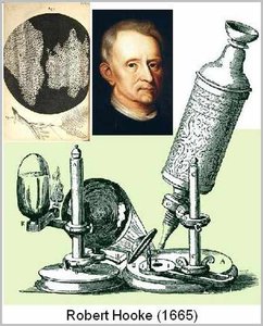
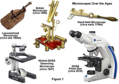
Microscopy has been essential for the development of cell biology. Early microscopes had limited resolution and resolving power, but technological improvements allowed scientists to observe finer details of cells and their components.
Resolution: The minimum distance two points must be apart to be distinguished as separate objects.
Resolving Power: The ability of a microscope to show fine detail.
Compound Microscope: Introduced in the 1830s, it uses two lenses for improved magnification and resolution, allowing visualization of structures as small as 1 micrometer.
The Cell Theory
The cell theory is a foundational concept in biology, developed in the 19th century by German scientists. It states that all living organisms are composed of cells, and all cells arise from preexisting cells.
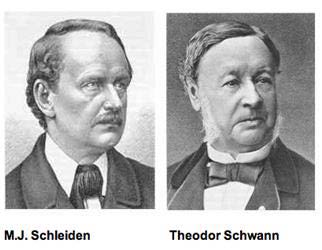
Matthias Schleiden and Theodor Schwann (1830s): Proposed that all organisms consist of one or more cells and that the cell is the basic unit of structure for all organisms.
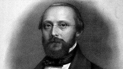
Rudolf Virchow (1850s): Added that all cells arise only from preexisting cells.
The Cell Theory Applies to All Organisms
The principles of cell theory are universal, applying to all forms of life, from bacteria to plants and animals. This universality underscores the importance of cells as the fundamental units of life.
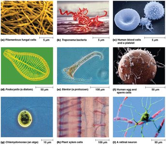
Modern Cell Biology: The Three Strands
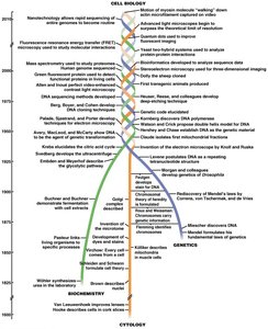
Modern cell biology is built upon three interwoven strands of scientific inquiry: cytology, biochemistry, and genetics. Each contributes unique perspectives and techniques to our understanding of cells.
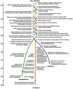
Cytology
Cytology focuses on the structure of cells and their components, utilizing various microscopy techniques to measure and visualize cellular dimensions and organelles.
Micrometer (μm): 10-6 meters; used for measuring cells.
Nanometer (nm): 10-9 meters; used for subcellular organelles.
Angstrom (Å): 0.1 nm; used for molecules like DNA, RNA, and proteins.
Microscopy Techniques in Cytology
Various types of microscopy have been developed to study cells and their structures:
Light Microscopy: The earliest tool for identifying large subcellular organelles.
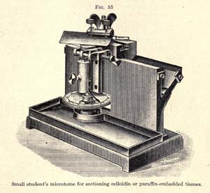
Microtome: Device for slicing very thin sections of samples for microscopy.
Special Optical Techniques: Include phase-contrast, differential interference contrast, fluorescence, and confocal microscopy for observing living cells.
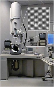
Electron Microscopy: Uses electron beams for much higher resolution and magnification than light microscopy.
Comparison of Light Microscopy Techniques
Type of Microscopy | Description | Application |
|---|---|---|
Brightfield (unstained specimen) | Passes light directly through specimen; little contrast unless stained | General cell observation |
Brightfield (stained specimen) | Staining enhances contrast but usually requires fixed (dead) cells | Detailed cell structure |
Phase contrast | Enhances contrast in unstained cells by amplifying variations in refractive index | Live cell imaging |
Differential interference | Uses optical modifications to exaggerate differences in refractive index | Live cell imaging |
Fluorescence | Shows locations of specific molecules using fluorescent dyes or proteins | Localization of proteins, organelles |
Confocal | Uses lasers and special optics for optical sectioning; sharp images of thin planes | 3D imaging of cells |
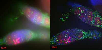
Brightfield vs. Confocal Microscopy
Confocal microscopy provides sharper, more detailed images than traditional brightfield microscopy, especially for thick specimens or those labeled with fluorescent markers.
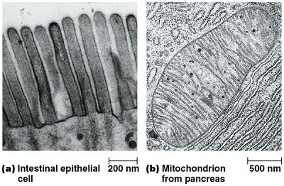
Electron Microscopy
Electron microscopy uses a beam of electrons instead of light, achieving up to 100,000x magnification and much higher resolution. There are two main types:
Transmission Electron Microscopy (TEM): Electrons pass through the specimen, revealing internal structures.
Scanning Electron Microscopy (SEM): Electrons scan the surface, providing detailed surface images.
Biochemistry
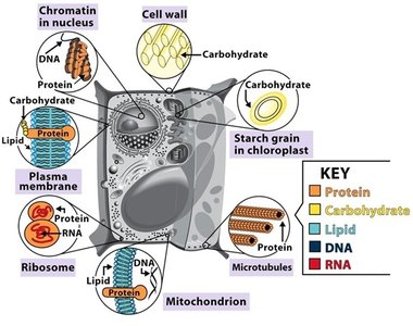
Biochemistry is the study of the chemical processes within living organisms. It focuses on the structure and function of biological molecules and the pathways of cellular reactions.
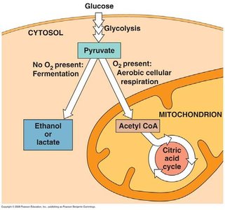
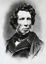
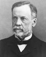
Historical Milestones: Synthesis of urea (Wöhler, 1828), fermentation by yeast (Pasteur, 1860s), and cell-free fermentation (Buchner, 1897).
Key Focus: Understanding the molecular basis of cellular structure and function.
Biochemical Methods
Subcellular Fractionation: Uses centrifugation to separate cellular components.
Ultracentrifuges: Spin at very high speeds (up to 100,000 rpm) to separate macromolecules.
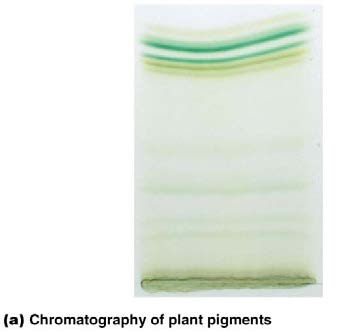
Chromatography: Separates molecules based on size, charge, or chemical affinity.
Electrophoresis: Uses an electric field to separate proteins, DNA, or RNA by size and charge.
Mass Spectrometry: Determines the size and composition of proteins.
Genetics
Genetics is the study of heredity and the flow of genetic information. It encompasses classical genetics, molecular genetics, and modern genomics.
Classical Genetics: Mendel's experiments with pea plants (1866) established the concept of hereditary factors (genes).
Chromosome Theory: Sutton and Boveri (1902) proposed that genes are located on chromosomes.
Molecular Genetics: Watson and Crick (1953) described the double helix structure of DNA, leading to the central dogma of molecular biology (DNA → RNA → Protein).
Genetic Information Flow
The central dogma of molecular biology describes the flow of genetic information within a cell:
DNA Replication: DNA makes a copy of itself.
Transcription: DNA is transcribed into RNA.
Translation: RNA is translated into protein.
Equation (Central Dogma):
Modern Genetic Techniques
Recombinant DNA Technology: Includes DNA cloning, transformation, and sequencing.
Bioinformatics: High-throughput analysis of genomes, transcriptomes, proteomes, metabolomes, and more.
Scientific Method and Model Systems
Cell biology relies on the scientific method and the use of model systems to test hypotheses and understand cellular processes.
Scientific Method: Systematic approach to inquiry involving observation, hypothesis formation, experimentation, and analysis.
Model Systems: Well-characterized species or cell cultures used for experimental studies (e.g., Drosophila melanogaster, yeast, mice).
Additional info: This summary integrates foundational concepts from the first chapter of a cell biology course, providing context for the development of the field and the essential tools and methods used in modern cell biology.
