 Back
BackBiological Membranes: Structure, Composition, and Function
Study Guide - Smart Notes
 Tailored notes based on your materials, expanded with key definitions, examples, and context.
Biological Membranes
Introduction to Membrane Fundamentals and Architecture
Biological membranes are essential for life, defining cellular boundaries and creating specialized compartments within cells. Their structure and function are central to understanding cellular physiology and pathology. Modern membrane biology integrates structural, biochemical, and clinical perspectives to elucidate the roles of membranes in health and disease.
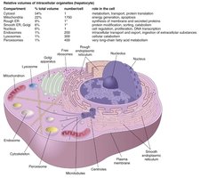
Cellular Boundaries: Membranes separate the cell from its environment and compartmentalize organelles.
Organelles with Membranes: Plasma membrane, nuclear envelope, endoplasmic reticulum, Golgi apparatus, mitochondria, lysosomes, and peroxisomes.
Selective Barrier Function
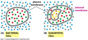
Cell membranes act as selective barriers, controlling the movement of substances into and out of the cell. In bacteria, the plasma membrane is the only membrane, while eukaryotes possess both plasma and internal membranes.
The Hydrophobic Core and Water Behavior
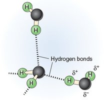
The hydrophobic core of the membrane is crucial due to the properties of water. Water is a dipolar molecule, forming transient hydrogen bonds that stabilize polar and charged solutes. Moving such solutes into the nonpolar membrane core is energetically unfavorable, making the bilayer an effective permeability barrier.
Hydration Shells: Polar/charged solutes are stabilized in water by hydration shells.
Energetic Cost: Removing hydration shells to cross the membrane is energetically costly.
Membrane Functions in Cellular Processes
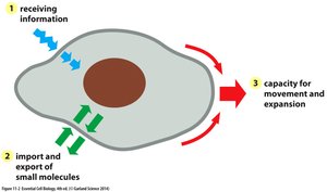
The plasma membrane is vital for cell communication, molecular transport, and cellular growth and motility. Membranes must facilitate the transfer of information and materials across their barriers.
Membrane Composition and Lipid Chemistry
Major Components of Biological Membranes
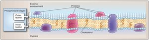
All biological membranes are composed of a lipid bilayer interspersed with proteins. The main lipid classes are phospholipids, cholesterol, and glycolipids.
Phospholipids: Amphipathic molecules with hydrophilic heads and hydrophobic tails, forming the bilayer structure.
Cholesterol: Modulates membrane fluidity and stability.
Glycolipids: Involved in cell recognition, signaling, and protection.
Phospholipid Structure and Amphipathicity
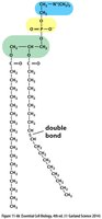
Phospholipids are the most abundant membrane lipids. They contain a glycerol backbone, two fatty acid tails (usually one saturated and one unsaturated), a phosphate group, and a variable head group (e.g., choline in phosphatidylcholine).
Amphipathic Nature: Hydrophilic head interacts with water; hydrophobic tails avoid water, driving bilayer formation.
Fatty Acids: Saturation and Fluidity
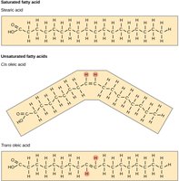
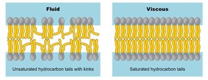
Fatty acids are long hydrocarbon chains that may be saturated (no double bonds) or unsaturated (one or more double bonds). The degree of saturation and chain length influence membrane fluidity.
Saturated Fatty Acids: Straight chains, pack tightly, decrease fluidity.
Unsaturated Fatty Acids: Kinked chains (cis double bonds), pack loosely, increase fluidity.
Chain Length: Longer chains increase van der Waals interactions, decreasing fluidity.
Cholesterol's Role in Membranes
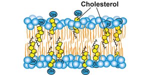
Cholesterol is intercalated among phospholipids, modulating membrane fluidity. At body temperature, it restricts phospholipid movement, decreasing fluidity. At lower temperatures, it prevents tight packing, maintaining fluidity.
Fluidity Buffer: Cholesterol narrows the range of membrane fluidity across temperatures.
Glycolipids and Glycoproteins
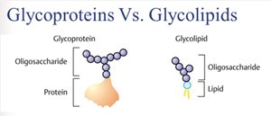
Glycolipids and glycoproteins are membrane components with attached carbohydrate chains. They play roles in cell recognition, signaling, and protection, and are clinically significant in cancer, immune response, and lysosomal storage diseases.
Membrane Dynamics and Organization
Lipid Bilayer Formation and Properties
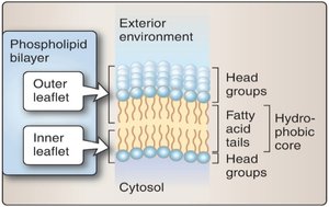
Amphipathic phospholipids spontaneously form bilayers in water, with hydrophobic tails facing inward and hydrophilic heads facing outward. This arrangement minimizes energetically unfavorable interactions with water.
Membrane Fluidity and Lipid Movement
Membrane fluidity is influenced by lipid composition and temperature. Phospholipids can move laterally, rotate, and flex, but rarely flip-flop between leaflets without enzymatic assistance.
Phosphatidylserine (PS) Externalization in Apoptosis
During apoptosis, PS is translocated from the inner to the outer leaflet of the plasma membrane, serving as an "eat me" signal for phagocytes. This process is tightly regulated and distinguishes apoptosis from necrosis.
Step 1: Apoptotic signal activates scramblase, inactivates flippase.
Step 2: PS externalizes to the outer leaflet.
Step 3: Phagocytes recognize externalized PS.
Step 4: Apoptotic cell is cleared without inflammation.
Membrane Proteins and Functional Specialization
Types and Roles of Membrane Proteins
Membrane proteins are responsible for most of the membrane's biological functions. They can be integral (spanning the bilayer), peripheral (associated with the membrane surface), or lipid-anchored.
Integral Proteins: Span the membrane, often as α-helices or β-barrels.
Peripheral Proteins: Attach to membrane surfaces, often via interactions with phospholipid head groups or integral proteins.
Lipid-Anchored Proteins: Covalently attached to lipids within the membrane.
Functions: Transport, signal transduction, cell adhesion, recognition, enzymatic activity, cytoskeletal attachment.
Membrane Carbohydrates and the Glycocalyx
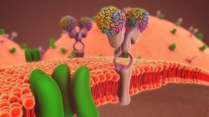
Membrane carbohydrates are covalently attached to lipids and proteins, forming the glycocalyx. This structure is involved in cell-cell recognition, adhesion, protection, and regulation of vascular and immune functions.
The Fluid Mosaic Model and Membrane Domains
Fluid Mosaic Model
The fluid mosaic model describes the membrane as a dynamic, asymmetric structure with laterally mobile components. Membrane asymmetry is established during synthesis in the ER and Golgi and is critical for function.
Key Principles: Dynamic structure, lateral movement, selective permeability, asymmetric organization.
Lipid Rafts and Membrane Microdomains
Lipid rafts are specialized membrane microdomains enriched in cholesterol, sphingolipids, and specific proteins. They serve as platforms for protein organization, signal transduction, and membrane trafficking. Disruption of rafts is implicated in diseases such as insulin resistance and viral entry (e.g., SARS-CoV-2).
Conclusion
Membrane abnormalities underlie many diseases.
Understanding membrane structure aids drug design and diagnostics.
Membrane targeting is a key therapeutic strategy.
