 Back
BackCell Adhesions, Cell Junctions, and Extracellular Structures: Study Notes
Study Guide - Smart Notes
 Tailored notes based on your materials, expanded with key definitions, examples, and context.
Beyond the Cell: Cell Adhesions, Cell Junctions, and Extracellular Structures
Introduction
This chapter explores how animal cells interact with each other and with the extracellular matrix (ECM) to form tissues and organs. It covers the structure and function of cell junctions, the composition and roles of the ECM, and the molecular mechanisms underlying cell adhesion and communication.
Types of Animal Tissues
Overview of Tissue Types
Epithelium: Sheets of polarized cells with distinct apical and basal domains. Functions include absorption, secretion, and protection.
Connective Tissue: Loosely organized cells embedded in abundant ECM, providing structural support and elasticity.
Muscle and Nervous Tissue: Specialized for contraction and signal transmission, respectively.
Both epithelial and connective tissues contain an ECM, but its abundance and importance vary by tissue type.
Cell Junctions
Overview of Cell Junctions
Cell junctions are specialized structures that connect adjacent cells or cells to the ECM, maintaining tissue integrity and function. Major types include:
Adhesive Junctions: Anchor the cytoskeleton to the cell surface and mediate cell-cell adhesion.
Tight Junctions: Seal adjacent cells to prevent the passage of molecules between them.
Gap Junctions: Allow direct communication between cells by forming channels for ions and small molecules.
Adhesive Junctions
Adherens Junctions: Cadherin-mediated, connect to actin filaments. Prominent in epithelial tissues.
Desmosomes: Button-like points of strong adhesion, connect to intermediate filaments. Abundant in tissues under mechanical stress (e.g., skin, heart).
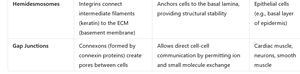
Hemidesmosomes: Anchor cells to the basal lamina via integrins.
Adhesive junctions are dynamic, assembling and disassembling in response to cellular events and signaling.
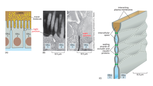
Tight Junctions
Tight junctions form a continuous seal around epithelial cells, preventing the movement of solutes between cells and maintaining distinct apical and basal membrane domains. Key proteins include claudins and occludins.
Gap Junctions
Gap junctions are formed by connexin proteins that assemble into connexons, creating direct channels between the cytoplasm of adjacent cells. They allow the passage of ions and small molecules, facilitating electrical and chemical communication, especially in cardiac and smooth muscle and neurons.
Summary Table: Major Cell Junctions
Junction Type | Main Components | Function | Location |
|---|---|---|---|
Hemidesmosomes | Integrins, intermediate filaments | Anchor cells to basal lamina | Epithelial cells (basal layer) |
Gap Junctions | Connexins | Direct cell-cell communication | Cardiac muscle, neurons |
Cell Adhesion Molecules
Cadherins
Cadherins: Calcium-dependent adhesion molecules mediating homophilic cell-cell interactions. Different types (E-cadherin, P-cadherin) are tissue-specific.
Structure: Extracellular domains interact in a zipper-like fashion; cytosolic tails bind to catenins, linking to the actin cytoskeleton.
Role in Development and Disease: Changes in cadherin expression are critical in processes like the epithelial-mesenchymal transition (EMT) and cancer metastasis.
Integrins
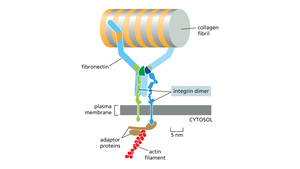
Integrins: Heterodimeric transmembrane receptors (α and β subunits) that mediate cell-ECM adhesion and signaling.
Binding: Bind ECM components such as fibronectin, laminin, and collagen.
Signaling: Involved in both "outside-in" and "inside-out" signaling, affecting cell migration, proliferation, and survival.
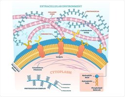
The Extracellular Matrix (ECM)
Definition and Functions
The ECM is a complex network of macromolecules secreted by cells, providing structural support, biochemical cues, and a scaffold for tissue organization. It is dynamic, constantly remodeled by cells.
Major Components of the ECM
Collagen: Most abundant protein in animals, provides tensile strength. Triple helix structure; many types (e.g., Type I in bone, Type IV in basal lamina).
Elastin: Provides elasticity, allowing tissues to stretch and recoil (e.g., in skin, lungs).
Proteoglycans and Glycosaminoglycans (GAGs): Hydrated, gel-like substances that resist compression. GAGs are long polysaccharides; proteoglycans are proteins with GAG chains attached.
Adhesive Glycoproteins: Fibronectin and laminin organize the ECM and mediate cell-ECM interactions.
Specialized ECM Structures
Basal Lamina: Thin, sheet-like ECM underlying epithelial cells and surrounding muscle, nerve, and fat cells. Composed of laminin, Type IV collagen, nidogen, and perlecan. Functions as a structural support and selective filter.
ECM Remodeling
Matrix Metalloproteinases (MMPs): Enzymes that degrade ECM components, facilitating tissue remodeling, wound healing, and cell migration.
Tissue Inhibitors of Metalloproteinases (TIMPs): Regulate MMP activity to maintain ECM integrity.
Summary Table: ECM Components and Functions
Component | Description | Function |
|---|---|---|
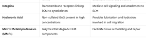
Integrins | Transmembrane receptors linking ECM to cytoskeleton | Mediate cell signaling and attachment to ECM |
Hyaluronic Acid | Non-sulfated GAG, high concentrations | Lubrication, hydration, cell migration |
Matrix Metalloproteinases (MMPs) | Enzymes degrading ECM | Tissue remodeling and repair |
Proteoglycans and Glycosaminoglycans (GAGs)
Structure and Function
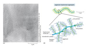
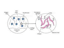
Proteoglycans: Core proteins with covalently attached GAG chains. Form hydrated gels that resist compression, especially in cartilage.
GAGs: Long, unbranched polysaccharides with repeating disaccharide units (e.g., chondroitin sulfate, keratan sulfate, hyaluronate).
Hyaluronate: Unique GAG that exists both as a free molecule and as a backbone for proteoglycan aggregates. Provides lubrication in joints.
Adhesive Glycoproteins
Fibronectin
Role: Bridges cells and ECM, essential for cell migration, wound healing, and tissue organization.
Clinical Relevance: Altered fibronectin expression is associated with cancer metastasis.
Laminin
Role: Major component of the basal lamina, influences cell differentiation, migration, and adhesion.
Integrins and Cell-ECM Interactions
Integrin Structure and Function
Structure: Heterodimers (α and β subunits) spanning the plasma membrane.
Function: Link ECM components (e.g., fibronectin, collagen) to the actin cytoskeleton via adaptor proteins. Mediate bidirectional signaling.
ECM and Disease
Examples of ECM-Related Diseases
Cancer: Altered ECM composition and increased MMP activity promote tumor invasion and metastasis.
Genetic Disorders: Mutations in collagen genes cause diseases like osteogenesis imperfecta and Ehlers-Danlos syndrome.
Fibrosis: Excessive ECM deposition leads to tissue scarring (e.g., liver cirrhosis).
Arthritis: Degradation of cartilage ECM impairs joint function.
Key Concepts and Applications
Cell adhesion and ECM interactions are essential for tissue structure, signaling, and function.
Disruption of cell junctions or ECM components can lead to disease.
Understanding these mechanisms is critical for fields such as developmental biology, pathology, and regenerative medicine.
