 Back
BackCell Biology: Structure, Function, and Membrane Dynamics
Study Guide - Smart Notes
 Tailored notes based on your materials, expanded with key definitions, examples, and context.
Cell Biology Overview
Definition and Discovery of the Cell
The cell is the fundamental unit of structure and function in all living organisms. Its discovery was enabled by the development of the microscope, which allowed scientists to observe details not visible to the naked eye.
Cell: Basic functional unit of structure and function in living organisms.
Discovery:
Microscopes revealed details invisible to the naked eye.
Robert Hooke observed cell walls in dead cork tissue.
Anton van Leeuwenhoek observed living cells (now called microorganisms).
Cell Theory
Principles of Cell Theory
Cell theory is a foundational concept in biology, describing the properties and functions of cells.
All organisms are composed of one or more cells.
The cell is the basic unit of structure and function in organisms.
All cells arise from pre-existing cells.
Cells contain hereditary information passed from cell to cell during division.
Cells as Basic Functional Units
Cells can exist as single units or as part of multicellular organisms, performing essential life functions.
Single-celled organisms: e.g., bacteria, protozoa.
Multicellular organisms: e.g., plants, animals.
Prokaryotic vs. Eukaryotic Cells
Cells are classified as prokaryotic or eukaryotic based on their structural features.
Prokaryotic Cells:
No nucleus.
No membrane-bound organelles.
Example: Bacteria.
Eukaryotic Cells:
Have a nucleus.
Contain membrane-bound organelles.
Examples: Plants, animals, fungi, protists.
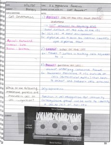
Cell Membrane and Permeability
Structure and Function of the Cell Membrane
The cell membrane is a selective barrier that regulates the passage of substances into and out of the cell.
Composed of a phospholipid bilayer with embedded proteins.
Cholesterol (in animal cells) stabilizes membrane fluidity.
Membrane proteins serve various functions, including transport, signaling, and structural support.
Phospholipid Bilayer
The phospholipid bilayer forms the basic structure of the cell membrane, providing both flexibility and selective permeability.
Each phospholipid has a hydrophilic head and hydrophobic tail.
Bilayer arrangement: Hydrophilic heads face outward, hydrophobic tails face inward.
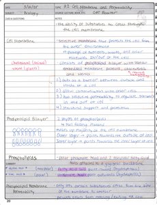
Membrane Permeability
Membrane permeability refers to the ability of substances to cross the cell membrane.
Small, nonpolar molecules (e.g., O2, CO2) pass easily.
Large or charged molecules require transport proteins.
Selective permeability is essential for maintaining cellular homeostasis.
Organelles and Cell Structures
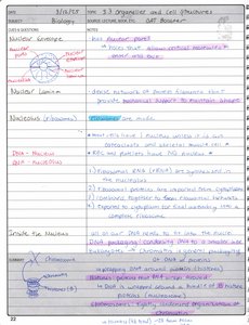
Nucleus
The nucleus is the control center of the cell, containing genetic material and regulating gene expression.
Surrounded by a nuclear envelope with pores for transport.
Contains nucleolus (site of ribosome synthesis).
Chromatin: DNA and associated proteins.
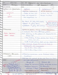
Cytoplasm vs. Cytosol
The cytoplasm includes all contents within the cell membrane except the nucleus, while the cytosol is the fluid portion.
Cytoplasm: Includes organelles and cytosol.
Cytosol: Jelly-like fluid containing dissolved substances.
Ribosomes
Ribosomes are the sites of protein synthesis, found either free in the cytosol or attached to the endoplasmic reticulum.
Composed of RNA and proteins.
Free ribosomes synthesize proteins for use within the cell.
ER-bound ribosomes synthesize proteins for secretion or membrane insertion.
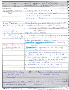
Endoplasmic Reticulum (ER)
The ER is a network of membranes involved in protein and lipid synthesis.
Rough ER: Studded with ribosomes; synthesizes proteins.
Smooth ER: Lacks ribosomes; synthesizes lipids and detoxifies chemicals.
Golgi Apparatus
The Golgi apparatus modifies, sorts, and packages proteins and lipids for transport.
Receives vesicles from the ER.
Processes and ships cellular products to their destinations.
Lysosomes and Peroxisomes
Lysosomes contain digestive enzymes for breaking down cellular waste, while peroxisomes detoxify harmful substances.
Lysosomes: Digest macromolecules, old organelles, and pathogens.
Peroxisomes: Break down fatty acids and detoxify hydrogen peroxide.
Mitochondria and Chloroplasts
Mitochondria are the powerhouses of the cell, generating ATP through cellular respiration. Chloroplasts, found in plant cells, conduct photosynthesis.
Mitochondria: Site of aerobic respiration; contain their own DNA.
Chloroplasts: Site of photosynthesis; contain their own DNA.
Membrane Proteins and Junctions
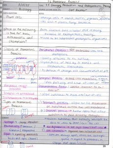
Classes of Membrane Proteins
Membrane proteins are classified based on their location and function.
Peripheral proteins: Not embedded in the membrane; attached to its surface.
Integral proteins: Embedded within the membrane; often span the bilayer.
Transmembrane proteins: Span the entire membrane; involved in transport and signaling.
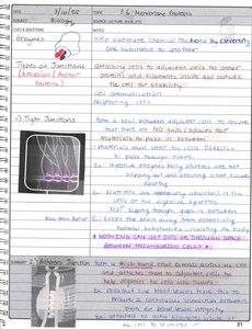
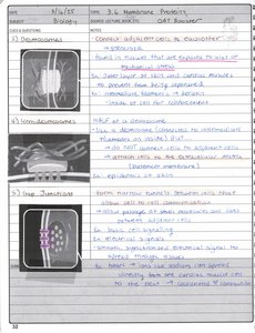
Membrane Junctions
Cells are connected by specialized junctions that regulate communication and adhesion.
Tight junctions: Seal adjacent cells, preventing leakage of molecules.
Desmosomes: Anchor cells together, providing mechanical strength.
Gap junctions: Allow direct communication between cells via channels.
Cell Transport
Passive and Active Transport
Cell transport mechanisms regulate the movement of substances across the membrane.
Passive transport: No energy required; includes diffusion and osmosis.
Active transport: Requires energy (ATP); moves substances against concentration gradients.
Equations:
Osmosis and diffusion are governed by concentration gradients:
Endocytosis and Exocytosis
Cells use endocytosis to engulf substances and exocytosis to expel them.
Endocytosis: Cell membrane engulfs material, forming a vesicle.
Exocytosis: Vesicle fuses with membrane, releasing contents outside.
Osmosis and Tonicity
Osmosis is the movement of water across a membrane, influenced by tonicity (relative solute concentration).
Hypertonic: Higher solute concentration outside; cell loses water.
Hypotonic: Lower solute concentration outside; cell gains water.
Isotonic: Equal solute concentration; no net water movement.
Cell Wall and Extracellular Matrix
Cell Wall
Plant cells and some bacteria have a cell wall for structural support and protection.
Composed of cellulose (plants) or peptidoglycan (bacteria).
Provides rigidity and prevents excessive water uptake.
Extracellular Matrix (ECM)
The ECM is a network of proteins and carbohydrates outside the cell, providing structural support and regulating cell behavior.
Major components: collagen, proteoglycans, fibronectin.
Functions: cell adhesion, signaling, and tissue integrity.
Cell Signaling and Movement
Cell Signaling
Cells communicate via molecular signals, which regulate growth, differentiation, and responses to the environment.
Signaling molecules bind to receptors, triggering intracellular pathways.
Examples: hormones, neurotransmitters.
Cytoskeleton and Cell Movement
The cytoskeleton is a network of protein filaments that provides structural support and enables cell movement.
Components: microtubules, microfilaments, intermediate filaments.
Functions: cell shape, intracellular transport, motility (e.g., cilia, flagella).
Energy, Evolution, and Endosymbiotic Theory
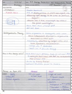
Energy Production
Cells generate energy through processes such as cellular respiration and photosynthesis.
Cellular respiration: Occurs in mitochondria; converts glucose to ATP.
Photosynthesis: Occurs in chloroplasts; converts light energy to chemical energy.
Endosymbiotic Theory
The endosymbiotic theory explains the origin of mitochondria and chloroplasts as formerly independent prokaryotes engulfed by ancestral eukaryotic cells.
Evidence: organelles have their own DNA, double membranes, and reproduce independently.
Plant vs. Animal Cells
Plant and animal cells share many features but differ in key structures.
Feature | Plant Cell | Animal Cell |
|---|---|---|
Cell Wall | Present | Absent |
Chloroplasts | Present | Absent |
Vacuole | Large central | Small or absent |
Centrioles | Absent | Present |
Summary Table: Cell Structures and Functions
Organelle | Function |
|---|---|
Nucleus | Genetic information storage, gene expression regulation |
Ribosome | Protein synthesis |
ER (Rough/Smooth) | Protein/lipid synthesis |
Golgi Apparatus | Modification, sorting, packaging of proteins/lipids |
Lysosome | Digestion of macromolecules |
Mitochondria | ATP production |
Chloroplast | Photosynthesis |
Cell Membrane | Selective permeability, protection |
Cell Wall | Structural support |
Cytoskeleton | Cell shape, movement |
Additional info: Academic context was added to expand brief points and ensure completeness. Diagrams were included only when directly relevant to the explanation.
